Advertisement
Advertisement
August 2026
Sponsored by Medtronic
Preserving Dialysis Fistula Patency With Arteriovenous DCB

Maintaining durable vascular access remains a central challenge in the care of patients requiring hemodialysis. Arteriovenous fistulas (AVFs) are generally preferred due to their lower infection risk and greater long-term durability compared with other access types. Stenosis caused by neointimal hyperplasia and adverse local hemodynamics frequently leads to access dysfunction, impaired dialysis delivery, and repeat reintervention.1 Plain old balloon angioplasty (POBA) remains the standard endovascular approach for treating symptomatic AVF stenosis, but restenosis after POBA is common, particularly in complex lesion locations such as the perianastomotic region, venous outflow, and cephalic arch.1 As a result, treatment approaches that may improve patency and reduce the frequency of repeat procedures are of substantial clinical interest.
Drug-coated balloons (DCBs) were developed to deliver an antiproliferative agent locally at the time of angioplasty, with the goal of inhibiting restenosis while avoiding a permanent implant. However, in hemodialysis access, the evidence base for DCBs has been relatively limited and heterogeneous. Compared with other vascular beds, there are few randomized controlled trials (RCTs) evaluating DCBs for dysfunctional AVFs specifically. Key prospective RCTs include the Lutonix AV trial, the IN.PACT AV Access investigational device exemption trial, and the investigator-led PAVE trial. Findings across these studies have not been uniform. The Lutonix AV trial showed modest improvement in target lesion primary patency at 1 year compared with POBA,2 whereas the PAVE trial did not demonstrate a patency advantage for Lutonix™* AV DCB (BD Interventional) treatment.3 In contrast, the IN.PACT AV Access RCT demonstrated significantly higher target lesion primary patency and fewer reinterventions with DCB compared with POBA at 6 and 12 months, with durability of the efficacy benefit reported through 36 months.4-6
Despite these data, prospective evidence remains limited, and RCT populations may not fully reflect routine practice. Real-world and post-approval registry data therefore provide complementary evidence across broader patient, access, and lesion characteristics.
The recently published multicenter, prospective IN.PACT AV post-approval study reported a 12-month target lesion primary patency rate of 70.2%, with a target lesion revascularization rate of 38.9%.7 At 12 months, the mean number of interventions required to maintain target lesion patency and access circuit patency was 0.6 ± 1.0 and 0.7 ± 1.1, respectively. The study also met its primary safety endpoint, with a 12-month serious infection rate of 13.4% against a 30% performance goal. These findings support the safety and effectiveness of DCB therapy beyond controlled trial settings.
Additional technologies, such as scoring or cutting balloons, may also be used to optimize lesion preparation before definitive therapy, particularly in resistant or recurrent stenoses. However, robust comparative data for these approaches in real-world AVF populations remain limited, and outcomes are highly dependent on lesion characteristics and procedural strategy.
The following case examples highlight the practical use of DCBs in common clinical scenarios encountered during routine dialysis access maintenance, including anastomotic stenosis in a radiocephalic AVF and recurrent cephalic arch stenosis in a brachiocephalic AVF.
CASE STUDY 1: ANASTOMOTIC STENOSIS IN A RADIOCEPHALIC AVF
Patient Presentation
A man in his mid 70s underwent left radiocephalic AVF creation in 2025 after previously requiring a tunneled central venous catheter (CVC) in the right internal jugular vein for urgent dialysis initiation. In early 2026, he was sent for a fistulogram due to difficult cannulation.
The patient was positioned on the angiography table with his left arm extended on an arm board. After the left forearm was prepped and draped in the usual sterile fashion, ultrasound was used to look for stenosis within the inflow and cannulation zone. The decision was made to access the fistula in a retrograde fashion to focus on the inflow.
Procedural Overview
After 4-F retrograde percutaneous micropuncture access and compression imaging confirming anastomotic stenosis, a stiff, angled 0.035-inch guidewire was directed into the proximal radial artery through a Berenstein catheter. The 4-F sheath was exchanged for a 6-F sheath. Imaging showed severe anastomotic stenosis (Figure 1A) and midforearm radial artery spasm.
A 3- X 100-mm standard POBA was performed within the mid/distal radial artery and AV anastomosis (Figure 1B). This was followed by 4- X 40-mm standard POBA to serially dilate the anastomosis. Finally, a 5- X 40-mm IN.PACT™ AV DCB (Medtronic) was inflated within the anastomosis for 3 full minutes (Figure 1C).
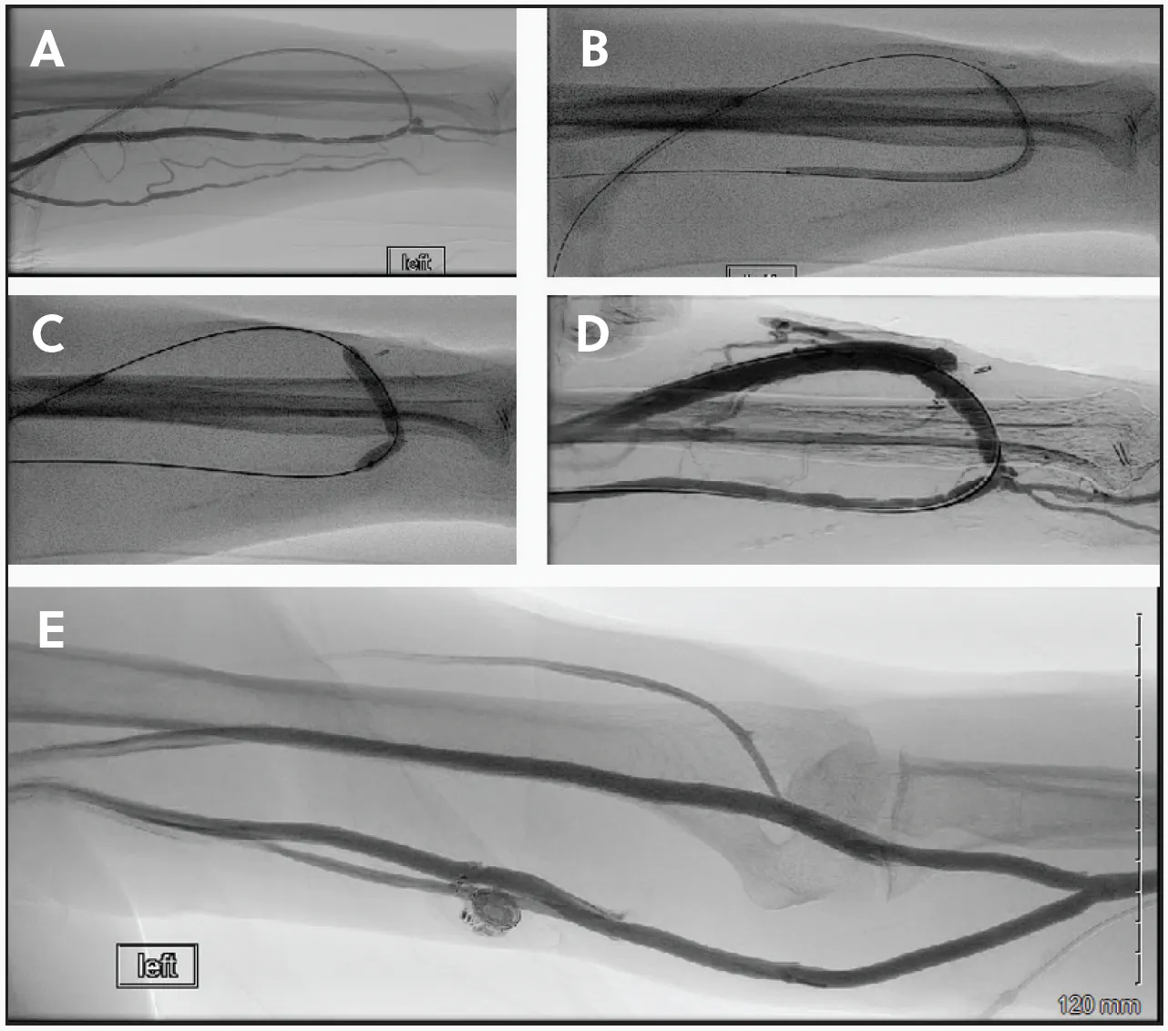
Figure 1. The initial left AVF fistulogram showing severe anastomotic stenosis (A). Standard POBA within the mid/distal radial artery and AV anastomosis (B). The 5- X 40-mm IN.PACT™ AV drug-coated balloon within the anastomosis (C). The final fistulogram showing the patent AVF (D). Outflow image showing wide patency of the upper arm outflow veins (E).
Procedural Results
Final fistulogram imaging showed wide patency of the treated segment, with no residual stenosis (Figure 1D). Outflow imaging showed wide patency of the upper arm outflow veins (Figure 1E) and central venous system. The patient’s CVC was removed about 1 month after successful cannulation and dialysis treatment via the AVF. At the time of writing, no issues had been reported with use of his access since the procedure.
CASE STUDY 2: RECURRENT CEPHALIC ARCH STENOSIS IN A BRACHIOCEPHALIC AVF
Patient Presentation
A man in his late 60s with a left brachiocephalic AV fistula created in 2019 presented for his first intervention in mid 2024, undergoing high-pressure balloon angioplasty followed by DCB angioplasty of the cephalic arch. The same procedure was performed a second time almost 6 months later. The patient was referred again approximately 9 months thereafter for enlarging pseudoaneurysms within the cannulation zone, prompting another fistulogram.
The patient was placed in the supine position on the angiography table with his left arm positioned on an arm board. The left upper arm was then prepped and draped in the usual sterile fashion. An ultrasound showed no concerns with inflow. A 4-F micropuncture access was placed antegrade. Compression imaging showed no inflow stenosis. Outflow imaging showed recurrent cephalic arch stenosis (Figure 2A).
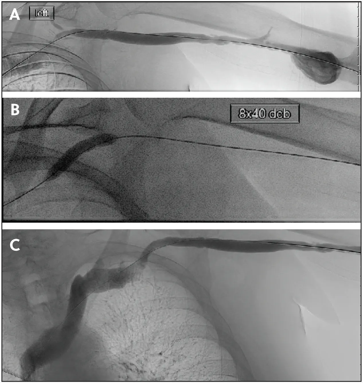
Figure 2. The initial left AVF fistulogram showing cephalic arch stenosis (A). The 8- X 40-mm IN.PACT AV DCB within the anastomosis (B). The final fistulogram showing improved patency (C).
Procedural Overview
A stiff 0.035-inch guidewire was placed centrally, and the 4-F sheath was exchanged for a 7-F sheath. A 7- X 60-mm standard POBA was performed within the cephalic arch lesion. Mild waisting was noted at burst pressure. A 7- X 40-mm high-pressure POBA was performed, with waisting relieved at 18 mm Hg inflation. This was followed by an 8- X 40-mm IN.PACT AV DCB, inflated to nominal pressure for a full 3 minutes (Figure 2B).
Final imaging showed improvement in flow, without residual stenosis (Figure 2C).
CLOSING REMARKS
These two cases illustrate the practical use of DCB angioplasty for maintaining AVF function. Adequate lesion preparation followed by prolonged DCB inflation restored luminal patency and improved access flow in both anastomotic and cephalic arch stenoses. Notably, the cephalic arch case showed a longer interval between interventions after DCB treatment (well within the Kidney Disease Outcomes Quality Initiative guidelines1), suggesting durable clinical benefit.
Although outcomes remain influenced by anatomy, lesion location, and prior intervention history, these examples support DCBs as a definitive treatment option for dysfunctional AVFs after appropriate lesion preparation.
1. Lok CE, Huber TS, Lee T, et al; National Kidney Foundation. KDOQI clinical practice guideline for vascular access: 2019 update. Am J Kidney Dis. 2020;75(4 suppl 2):S1-S164. Published correction appears in Am J Kidney Dis. 2021;77:551. doi: 10.1053/j.ajkd.2019.12.001
2. Trerotola SO, Saad TF, Roy-Chaudhury P; Lutonix AV clinical trial investigators. The Lutonix AV randomized trial of paclitaxel-coated balloons in arteriovenous fistula stenosis: 2-year results and subgroup analysis. J Vasc Interv Radiol. 2020;31:1-14.e5. doi: 10.1016/j.jvir.2019.08.035
3. Karunanithy N, Robinson EJ, Ahmad F, et al. A multicenter randomized controlled trial indicates that paclitaxel-coated balloons provide no benefit for arteriovenous fistulas. Kidney Int. 2021;100:447-456. doi: 10.1016/j.kint.2021.02.040
4. Lookstein RA, Haruguchi H, Ouriel K, et al; IN.PACT AV access investigators. Drug-coated balloons for dysfunctional dialysis arteriovenous fistulas. N Engl J Med. 2020;383:733-742. doi: 10.1056/NEJMoa1914617
5. Holden A, Haruguchi H, Suemitsu K, et al; IN.PACT AV Access investigators. IN.PACT AV Access randomized trial: 12-month clinical results demonstrating the sustained treatment effect of drug-coated balloons. J Vasc Interv Radiol. 2022;33:884-894.e7. doi: 10.1016/j.jvir.2022.03.606
6. Lookstein R, Haruguchi H, Suemitsu K, et al; IN.PACT AV Access investigators. IN.PACT AV Access randomized trial of drug-coated balloons for dysfunctional arteriovenous fistulae: clinical outcomes through 36 months. J Vasc Interv Radiol. 2023;34:2093-2102.e7. doi: 10.1016/j.jvir.2023.07.007
7. Misra S, Atkinson C Jr, Dermody M, et al; IN.PACT AV Access post-approval investigators. IN.PACT AV Access post-approval study of a paclitaxel drug-coated balloon for the treatment of dysfunctional arteriovenous dialysis circuits. J Vasc Interv Radiol. 2026;37:108869. doi: 10.1016/j.jvir.2026.108869
Disclosures
Dr. Dermody: Consultant to Boston Scientific and Medtronic.
The device used in this study is commercialized under the name IN.PACT AV™ drug-coated balloon (DCB). The IN.PACT AV DCB is not available for sale outside the United States of America, Canada, or Japan. Outside of the United States, Canada, and Japan, the IN.PACT™ Admiral™ DCB is indicated for the treatment of failing arteriovenous access fistulas in patients with end-stage renal disease undergoing dialysis.
IN.PACT™ AV Paclitaxel-coated PTA balloon catheter
Brief Statement
Indications for Use
The IN.PACT™ AV paclitaxel-coated PTA balloon catheter is indicated for percutaneous transluminal angioplasty, after appropriate vessel preparation, for the treatment of obstructive lesions up to 100 mm in length in the native arteriovenous dialysis fistulae with reference vessel diameters of 4 to 12 mm.
Contraindications
The IN.PACT™ AV DCB is contraindicated for use in:
- Coronary arteries, renal arteries, and supra-aortic/cerebrovascular arteries
- Patients who cannot receive recommended antiplatelet and/or anticoagulant therapy
- Patients judged to have a lesion that prevents complete inflation of an angioplasty balloon or proper placement of the delivery system
- Patients with known allergies or sensitivities to paclitaxel
- Women who are breastfeeding, pregnant, or are intending to become pregnant, or men intending to father children. It is unknown whether paclitaxel will be excreted in human milk and whether there is a potential for adverse reaction in nursing infants from paclitaxel exposure.
Warnings
- Do not use air or any gaseous medium to inflate the balloon. Use only the recommended inflation medium (equal parts contrast medium and saline solution).
- Do not move the guidewire during inflation of the IN.PACT™ AV DCB.
- Do not exceed the rated burst pressure (RBP). The RBP is based on the results of in vitro testing. Use of pressures higher than RBP may result in a ruptured balloon with possible intimal damage and dissection.
- The safety of using multiple IN.PACT™ AV DCBs with a total drug dosage exceeding 15,105 µg paclitaxel has not been evaluated clinically.
Precautions
- Assess risks and benefits before treating patients with a history of severe reaction to or inability to tolerate contrast agents. Identify allergic reactions to contrast media and antiplatelet therapy before treatment and consider alternatives for appropriate management prior to the procedure.
- Administer appropriate drug therapy to the patient according to standard protocols for PTA before insertion of the dilatation catheter.
- Do not rinse or wipe the IN.PACT™ AV DCB catheter.
- Handle the product with caution to avoid any damage to the balloon coating or folded balloon.
- This product is not intended for the expansion or delivery of a stent or graft.
- Do not use the IN.PACT™ AV DCB for pre-dilatation or for post-dilatation.
- Do not expose the product to organic solvents such as alcohol.
- To reduce the potential for vessel damage, the inflated diameter of the balloon should approximately match the inner diameter of the vessel just distal to the lesion.
Potential Adverse Events
The potential adverse events associated with use of the device include but are not limited to: Abrupt vessel closure, allergic reaction, arrhythmias, arterial or venous aneurysm, arterial or venous thrombosis, death, dissection, embolization, hematoma, hemorrhage, hypotension/hypertension, infection, ischemia or infarction of tissue/organ, loss of permanent access, pain, perforation or rupture of the artery or vein, pseudoaneurysm, restenosis of the dilated vessel, shock, stroke, and vessel spasms or recoil.
Potential adverse events not captured above that may be unique to the paclitaxel drug coating include, but are not limited to: allergic/immunologic reaction, alopecia, anemia, gastrointestinal symptoms, hematologic dyscrasia (including leucopenia, neutropenia, thrombocytopenia), hepatic enzyme changes, histologic changes in vessel wall, including inflammation, cellular damage, or necrosis, myalgia/arthralgia, myelosuppression, and peripheral neuropathy. Refer to the Physician’s Desk Reference for more information on the potential adverse effects observed with paclitaxel.
CAUTION: Federal (USA) law restricts this device to sale by or on the order of a physician.
Important information: Indications, contraindications, warnings and instructions for use can be found in the product labeling supplied with each device, at www.medtronic.com/manuals or contact a Medtronic representative.
US-CV-2601756 ©2026 Medtronic. Medtronic, Medtronic logo are trademarks of Medtronic. ”TM* third party brands are trademarks of their respective owner. All other brands are trademarks of Medtronic. For global distribution. 08/2026
Advertisement
Advertisement