Advertisement
Advertisement
August 2026
The Dos and Don’ts of AV Access Thrombectomy
Practical approaches to a thrombosed arteriovenous dialysis access, from systematic evaluation and patient selection to procedural judgment and complication management.
Arteriovenous (AV) access thrombectomy remains one of the most common urgent procedures in the care of patients with end-stage kidney disease. Successful and timely management of a thrombosed access prevents complications related to missed dialysis treatments, hospitalizations, central venous catheter placement, and permanent access loss. Durable restoration of access function requires thoughtful patient selection, careful procedural planning, and systematic identification and management of any associated stenotic lesions contributing to thrombosis. This article focuses on practical considerations for mechanical percutaneous (endovascular) thrombectomy of AV dialysis access in the outpatient setting.
PREPROCEDURAL EVALUATION
A thrombosed access is rarely entirely unpredictable; clinical and dialysis-related indicators frequently precede access failure and may provide important clues regarding the location and mechanism of dysfunction. Information obtained from the dialysis unit can be particularly useful. Progressive difficulty with cannulation, prolonged bleeding after needle removal, worsening dialysis adequacy, laboratory values suggestive of recirculation, high venous pressures, or decreased blood flow rates may suggest progressive outflow stenosis prior to thrombosis. An immature, recently placed access with difficult cannulation, high arterial pressures, low blood flow rates, or decreased dialysis adequacy may indicate inflow stenosis. Additionally, dialysis flow sheets should be reviewed to identify hypotensive episodes that could be contributing to thrombosis.
A clear understanding of the access circuit prior to intervention improves procedural efficiency and reduces the likelihood of incomplete treatment or complications. The AV access circuit begins and ends in the heart, and successful thrombectomy requires addressing four key components: (1) thrombus removal; (2) “pullback” angiography to evaluate the access, venous outflow, and central veins; (3) angioplasty of any associated stenosis; and (4) dislodgement of the arterial plug.
In AV grafts, thrombosis most commonly results from venous outflow stenosis, typically at the graft-vein anastomosis. In AV fistulas (AVFs), the distribution of pathology is variable and may include juxta-anastomotic lesions, cephalic arch stenosis, swing segment narrowing, or multiple stenosis throughout the body of the fistula.
Inflow lesions are a common cause of early access failure and, if identified within the first 4 to 6 weeks postoperatively, should ideally be managed by the proceduralist who placed the access. These often reflect a correctable structural problem that may be more appropriately addressed surgically. In addition, manipulation of fresh anastomoses risks disruption of incompletely healed suture lines.
PATIENT SELECTION
Not all thrombosed accesses are suitable for percutaneous thrombectomy. Access infection is an absolute contraindication to endovascular procedures. Pain, erythema, localized tenderness, purulent drainage, or systemic symptoms should raise concern for superimposed infection, particularly in thrombosed grafts. In such circumstances, thrombectomy may precipitate bacteremia or septic embolization, and surgical management is preferred.
Large aneurysmal fistulas (mega fistulas) are a relative contraindication to outpatient endovascular thrombectomies, as they may have extensive clot burden (Figure 1) and embolic consequences will be more clinically significant. In such cases, temporary catheter placement followed by inpatient surgical thrombectomy may be a safer and more appropriate strategy than immediate endovascular intervention.
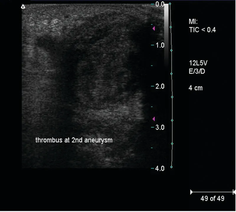
Figure 1. Extensive thrombus burden in an aneurysmal AVF.
Patient-specific physiologic considerations are equally important. Thrombectomy invariably results in some degree of pulmonary embolization. Although most patients tolerate this without clinical consequence, additional caution should be exercised in individuals with poor cardiopulmonary reserve, pulmonary hypertension, right ventricular dysfunction, or known right-to-left intracardiac shunts.
PROCEDURAL DESCRIPTION
If available, a preprocedural ultrasound is particularly valuable in thrombosed fistulas with uncertain anatomy or extensive aneurysmal degeneration. If the patient has had multiple previous accesses, the current thrombosed access should be marked preoperatively.
The first step is to obtain antegrade and retrograde access, ensuring there is adequate separation between the puncture sites to allow for thrombus removal from the entire access (Figure 2). For the “lyse and wait” technique, a thrombolytic agent may be injected along with a partially inflated angioplasty balloon to dam the outflow and allow the thrombolytic to dwell within the access, followed by aspiration of the thrombus. Once you have removed as much of the thrombus as possible, the next step is to gain access into the central venous system through the outflow. A straight catheter is then inserted over the wire, ensuring the wire is always ahead of the catheter. Contrast is then injected through the catheter to confirm patency of the central veins, administer any systemic medications, and then “pull back” to identify the site of stenosis (Figure 3), which should then undergo angioplasty (Figure 4). In a graft, the entire length of the graft undergoes angioplasty, but in a fistula, angioplasty should be limited to the specific stenotic segment. The inflow is then addressed, and the thrombotic plug is removed with an embolectomy (Figure 5). It is critical to partially deflate the catheter when passing through the anastomosis to avoid rupturing the anastomosis. If access flow remains sluggish, an antegrade arteriogram should be obtained. A retrograde arteriogram, although technically easier, increases the risk of distal clot embolization. The arteriogram allows for evaluation and management of the inflow artery, anastomosis, and juxta-anastomotic area. A final angiogram is then obtained to evaluate the entire circuit and address any residual or elastic lesions. It is optimal to have a soft thrill throughout the access at the end of the procedure, with adequate blood flow to support dialysis at the prescribed rate. Care should be taken to aim for procedural success rather than radiographic perfection.
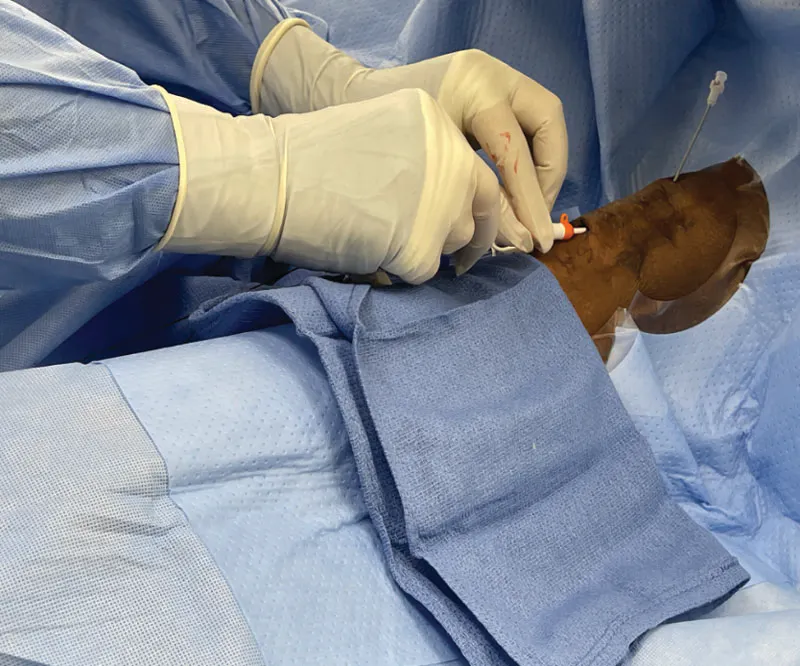
Figure 2. Antegrade and retrograde access, ensuring there is adequate separation between the two puncture sites
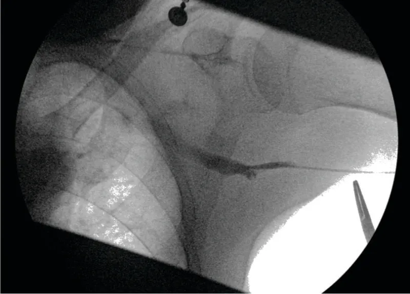
Figure 3. Pullback through 4-F catheter to localize site of stenosis.
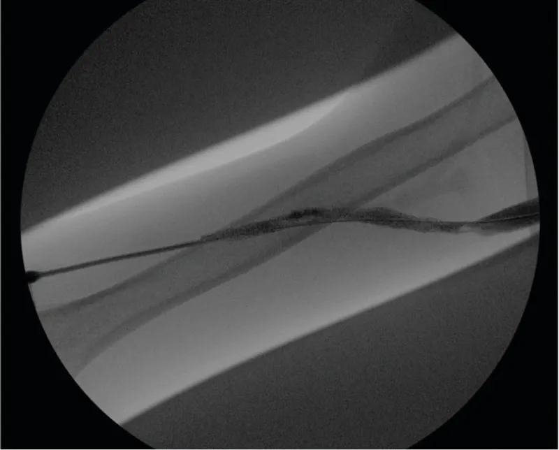
Figure 4. Outflow stenosis with residual thrombus.
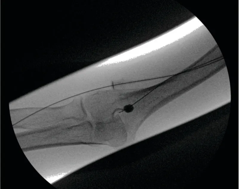
Figure 5. Embolectomy (Fogarty) balloon to dislodge clot at arterial anastomosis.
COMPLICATIONS
Although thrombectomy is generally safe and effective in experienced hands, complications remain unavoidable in a subset of cases. Procedural success depends not only on technical skill but also on anticipating complications before they occur and recognizing when continued intervention may no longer serve the patient’s best interest.
The most common complications are bleeding and hematoma formation, as our patients often have platelet dysfunction and receive intraprocedural anticoagulation. Excessive compression should be avoided during postprocedure hemostasis, as overly aggressive pressure may reocclude the access. If a suture is placed, it should be removed in 24 to 48 hours.
AV Access Thrombectomy Dos and Don’ts
General Considerations
- Determine why the access thrombosed before intervening, and incorporate dialysis unit data and physical examination into procedural planning.
- Remember that careful patient selection and addressing modifiable risk factors are key to a successful outcome.
- Evaluate the entire access circuit, and address all associated stenoses, including inflow, outflow, and central lesions.
- Don’t repeat thrombectomy reflexively without reassessing the long-term access strategy.
Technical Considerations
- Ensure adequate spacing between antegrade and retrograde sheaths to ensure complete thrombus removal.
- Always ensure the wire stays ahead of any catheters/balloons throughout the procedure.
- Partially deflate the embolectomy catheter when crossing the arterial anastomosis.
- Avoid retrograde arteriograms to assess inflow.
- Veins are forgiving; arteries are not.
- The enemy of good is better. Success is defined by reestablishing adequate blood flow to achieve dialysis prescription rather than radiographic perfection.
Vessel rupture represents another important consideration. Venous ruptures can frequently be managed conservatively with prolonged balloon tamponade and careful observation. However, arterial injury is associated with greater procedural consequences and underscores the importance of meticulous inflow manipulation. A useful procedural principle is worth emphasizing: Veins are forgiving, arteries are not.
Distal or proximal clot embolization can be limb- or life-threatening. Addressing patient-specific risk factors and procedural manipulations as described previously may minimize the risk of clinically significant emboli. Massive pulmonary emboli, although rare, require aggressive management, including supplemental oxygen, systemic anticoagulation, and embolectomy. Distal clot embolization into hands/fingers leading to ischemia may be managed by back bleeding, embolectomy, or thrombolytics.
RECURRENT THROMBOSIS AND ACCESS SUCCESSION PLANNING
Thrombectomy should never be viewed as an isolated procedure. Each intervention occurs within the broader context of a patient’s overall access trajectory and long-term dialysis plan. Recurrent thrombosis, particularly when occurring at increasingly short intervals, warrants reassessment rather than reflexive repeat intervention. The most effective access operators balance procedural success with preservation of future access options, protection of venous real estate, and thoughtful long-term planning.
CONCLUSION
Successful AV access thrombectomy requires substantially more than technical clot removal. Durable outcomes depend on thoughtful patient selection, systematic evaluation of the access circuit, careful treatment of the lesion responsible for thrombosis, and procedural judgment grounded in an understanding of access physiology. Although devices and techniques continue to evolve, many of the principles underlying successful thrombectomy remain unchanged: Understand why the access failed, treat the underlying lesion, proceed deliberately, and recognize when restraint is warranted. Ultimately, the goal of thrombectomy goes beyond simply restoration of patency; it includes safe and reliable return to dialysis, while preserving future access options whenever possible.
RECOMMENDED READING
Falk A, Trerotola SO, Valji K. Addressing the clotted access. Endovasc Today. 2013;12:52-54, 56, 58. https://evtoday.com/articles/2013-june/addressing-the-clotted-access
Lok CE, Huber TS, Lee T, et al; National Kidney Foundation. KDOQI clinical practice guideline for vascular access: 2019 update. Am J Kidney Dis. 2020;75(4 suppl 2):S1-S164. Published correction appears in Am J Kidney Dis. 2021;77:551. doi: 10.1053/j.ajkd.2019.12.001.
Niyyar VD, Work J. Interventional nephrology: core curriculum 2009. Am J Kidney Dis. 2009;54:169-82. doi: 10.1053/j.ajkd.2009.03.011
Trerotola SO, Scheel PJ Jr, Powe NR, et al. Screening for dialysis access graft malfunction: comparison of physical examination with US. J Vasc Interv Radiol. 1996;7:15-20. doi: 10.1016/s1051-0443(96)70726-1
Advertisement
Advertisement