Advertisement
Advertisement
July 2026
Sponsored by InterVene, Inc.
From Occluded to Open: How Recana® Is Restoring Flow in Complex Venous Disease
The Recana® Thrombectomy Catheter System represents a novel strategy for treating in-stent restenosis and residual native vein obstructions.
With Abdullah Shaikh, MD; Mr. Taha Khan; and Prof. Stephen Black
Deep venous stenting has increasingly been used as a treatment strategy for postthrombotic syndrome (PTS) and as an adjunctive therapy in acute deep vein thrombosis (DVT) interventions. The recently published C-TRACT data showed that in patients with moderate or severe PTS and iliac vein obstruction, endovascular therapy led to less severe PTS and better health-related quality of life than standard care over a 6-month period.1 The recent surge in mechanical thrombectomy interventions for acute DVT has resulted in the rapid rise of adjunctive venous stenting, with rates between 44% and 80%.2,3 In-stent restenosis (ISR) is a common complication of venous stenting, with up to 89% of stents having some degree of ISR at 12 months.4 Despite antiplatelet and anticoagulation regimens, ISR remains a significant complication, arising in both the early postprocedural period and later chronic phases.5-7 ISR develops through two principal mechanisms: (1) mesenchymal ingrowth through the stent mesh producing an extracellular matrix that narrows the vessel lumen, and (2) thrombus formation leading to progressive intimal thickening, a process that evolves over time from fresh and organized thrombus toward dense, fibrotic, and ultimately calcified material.8
In the setting of organized or chronic ISR, treatment options remain limited. Catheter-directed thrombolysis demonstrates poor efficacy once thrombus has matured beyond 21 days, with no successful recanalization reported in stents occluded beyond this threshold.9 Aspiration-based thrombectomy is similarly limited by the dense, fibrotic composition of chronic material. Venoplasty and stent relining do not remove existing thrombotic burden and provide only temporary luminal reshaping, with median luminal gains of approximately 31% to 42%.10
The Recana® Thrombectomy Catheter System (InterVene, Inc.) is a purpose-built, integrated platform designed to address the treatment gaps associated with ISR and improve outcomes for this challenging complication of venous stenting. The system features a debulking catheter with an expandable, helical coring element with a sharpened beveled edge; self-expanding nitinol baskets for material capture; and introducer and collection sheaths (Figure 1). The Recana System offers physicians a complete portfolio of devices designed to address the full spectrum of ISR and native vein occlusion complexity.
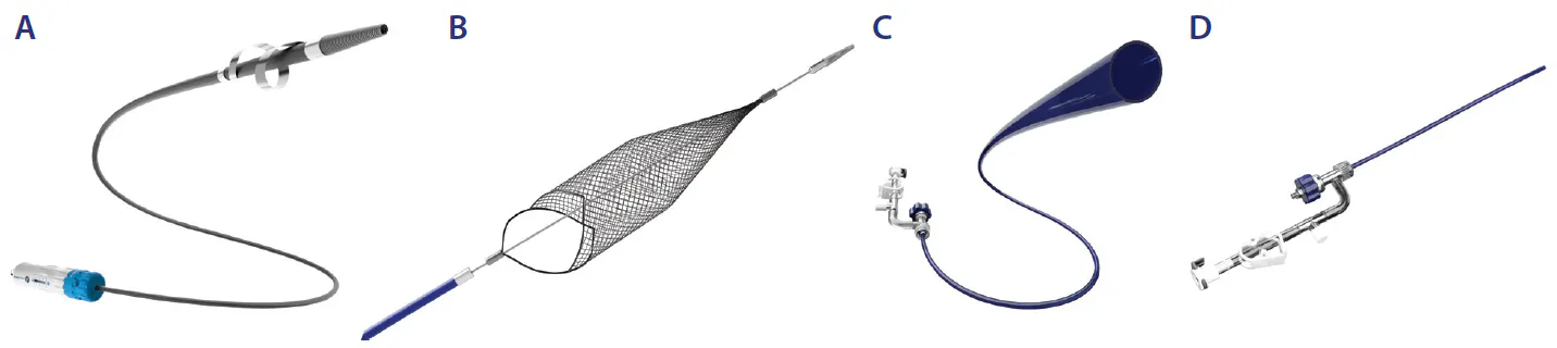
Figure 1. The Recana Thrombectomy Catheter System consists of a debulking catheter (A) with an expandable, helical coring element with a sharpened beveled edge; self-expanding nitinol baskets for material capture (B); a collection sheath (C); and introducer sheath (D).
InterVene recently announced the commencement of the Recana for Lower Extremity Venous Obstructions and In-Stent Restenosis (CALIBER) registry (NCT07529756).11 This prospective, multicenter, observational registry aims to gather safety and effectiveness data on the Recana System across a broad population of patients with symptomatic lower extremity venous obstructions and ISR. The CALIBER registry is currently enrolling patients across multiple centers in the United States, with results expected to inform best practices for the endovascular management of residual venous obstructions and ISR.
The following case studies describe patients with ISR and ongoing symptoms, highlighting the clinical decision-making, endovascular strategy, and real-world challenges encountered in managing significant ISR and inflow disease due to residual venous obstructions.
CASE 1: TREATING ISR AND NATIVE VESSEL OBSTRUCTION WITH THE RECANA SYSTEM

PATIENT PRESENTATION
A man in his mid-30s with prior DVT and ISR was evaluated 18 months prior for right leg symptoms. CT venography showed an atretic infrarenal inferior vena cava (IVC) and chronic thrombosis of the right common iliac (CIV) and external iliac veins (EIV) (Figure 1A). The patient reported moderate right leg pain, cramping, and heaviness that worsened at the end of his workdays. He remained compliant with anticoagulation and compression therapy. Due to the symptom impact to his profession, the decision for intervention with the Recana Thrombectomy Catheter System was made.
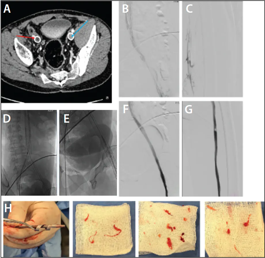
Figure 1. CT venography showing an occluded right EIV stent and patent left EIV stent (A). Little flow through stent with reflux into distal collaterals (B). Significant chronic obstruction in femoropopliteal vein (C). Recana collection basket deployed in IVC stent (D). Recana debulking catheter (E). Post-Recana robust flow with greater luminal area (F). Post-Recana native vein flow restored (G). Material captured and removed (H).
PROCEDURAL OVERVIEW
Right popliteal vein access was obtained under ultrasound guidance. The superficial femoral vein and stent were crossed with a 0.035-inch angled Glidewire (Terumo Interventional Systems) and catheter. Intravascular ultrasound (IVUS) confirmed intraluminal wire position. The left popliteal vein was then accessed using the same technique.
Both popliteal sheaths were exchanged for 16-F Recana introducer sheaths. An 18-mm nitinol Recana collection basket was advanced over the guidewire and deployed in the IVC stent to capture debulked material (Figure 1D).
The 13-F Recana debulking catheter was advanced through the introducer sheath for mechanical debulking within the right CIV and EIV stent (Figure 1E). The Recana debulking catheter's coring element was then expanded in diameter with a retrograde and antegrade scrubbing motion to debulk the occlusive material. A similar technique was performed in the native femoral vein (FV) to debulk the occlusive material and improve inflow.
CONCLUSION
The Recana System successfully treated ISR and restored flow in the native vein, thus improving inflow into the stent. Recana’s helical coring element allowed for directional debulking of occlusive material that was subsequently captured in the nitinol collection baskets placed distally during thrombectomy.
CASE 2: RESTORING PATENCY IN LONG-STANDING OCCLUSIVE ISR WITH THE RECANA SYSTEM


PATIENT PRESENTATION
A man in his early 40s with a history of DVT and PTS presented with moderate to severe leg swelling and pain. The patient underwent recanalization attempts over the years, including venous stent placement, balloon venoplasty, and stent relining, with only limited pain relief. Ultrasound demonstrated significant disease in the left iliac vein. The right side had significant scarring and FV disease but was not symptomatic. Intervention with the Recana System was chosen due to the patient’s history of failed treatments.
PROCEDURAL OVERVIEW
Bilateral venous access was gained in the left and right FVs using a 16-F DrySeal sheath (Gore & Associates). Right internal jugular (IJ) access was also obtained to allow for through-and-through access in the left FV. The Recana 18-mm nitinol collection basket was placed contralaterally in the IVC stent to capture debulked material. A 0.035-inch Amplatz super stiff wire (Boston Scientific Corporation) was introduced and flossed through the occlusion to the right IJ. Imaging confirmed a fully occluded venous stent with obstructive material visible in the CIV, EIV, and FV (Figure 1). The Recana debulking catheter was inserted over the wire, and the coring element was slowly expanded within the stent (Figure 2). With each successive pass, the Recana System advanced increasingly smoother through the lesion. Upon removal, a substantial amount of material was collected on the coring element (Figure 3). Adjunctive balloon angioplasty was then performed. Postprocedure imaging showed improved cephalad flow and increased luminal area (Figure 4).
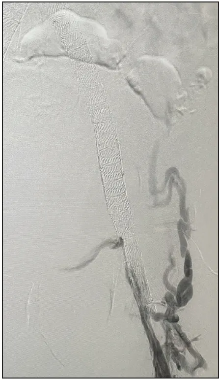
Figure 1. Preprocedure image confirming a fully occluded venous stent.
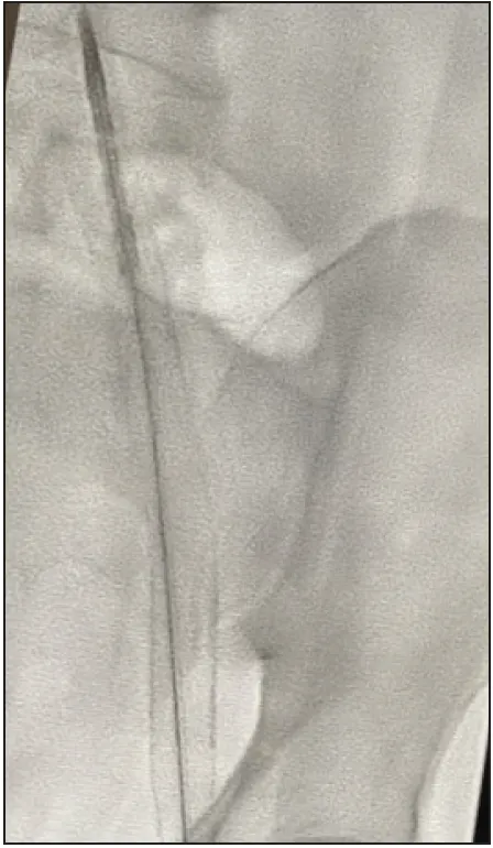
Figure 2. Insertion of the Recana debulking catheter.
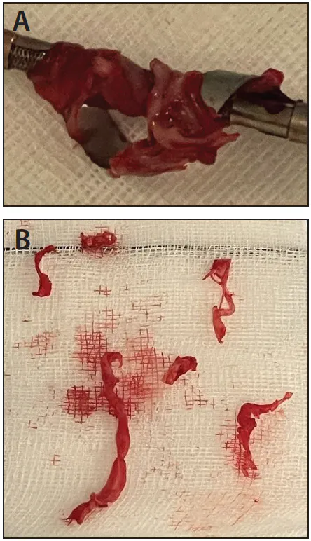
Figure 3. Material captured by the Recana System (A, B).
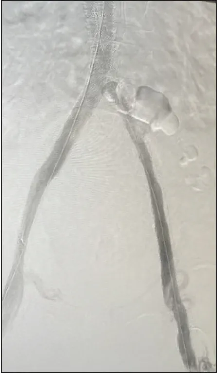
Figure 4. Postprocedure image showing increased luminal area.
CONCLUSION
The Recana System successfully restored flow and increased lumen area through the removal of obstructive ISR. This case highlights the versatility of the system, which can be used in both contralateral and ipsilateral approaches depending on patient condition and operator preference. The substantial volume and composition of the material removed shows how effective mechanical debulking can be for patients who have failed other therapies. Furthermore, the use of the collection basket for distal material capture proved to be a critical component in this case.
1. Vedantham S, Kahn SR, Marston WA, et al. Endovascular therapy for post-thrombotic syndrome–a randomized trial. N Engl J Med. Published online April 13, 2026. doi: 10.1056/NEJMoa2519001
2. Dexter D, Kado H, Shaikh A, et al. Safety and effectiveness of mechanical thrombectomy from the fully enrolled multicenter, prospective CLOUT registry. J Soc Cardiovasc Angiogr Interv. 2023;2:100585. doi: 10.1016/j.jscai.2023.100585
3. Gonzalez-Urquijo M, Vargas JF, Marine L, et al. Computer-assisted vacuum thrombectomy using Lightning 12 and Lightning Flash 16 in acute proximal lower extremity deep vein thrombosis. Phlebology. Published online November 26, 2025. doi: 10.1177/02683555251404147
4. Jayaraj A, Walker W, Raju S. In-stent restenosis after iliocaval stenting—characteristics and outcomes [abstract]. J Vasc Surg Venous Lymphat Disord. 2019;7:301-302. https://doi.org/10.1016/j.jvsv.2019.01.038
5. Aurshina A, Ascher E, Haggerty J, et al. Etiology of iliocaval stent thrombosis. J Vasc Surg Venous Lymphat Disord. 2020;8:207-210. doi: 10.1016/j.jvsv.2019.09.017
6. Sebastian T, Spirk D, Engelberger RP, et al. Incidence of stent thrombosis after endovascular treatment of iliofemoral or caval veins in patients with the postthrombotic syndrome. Thromb Haemost. 2019;119:2064-2073. doi: 10.1055/s-0039-1697955
7. Fu J, Tang B, Wang H, Luo H. Stent characteristics of 32 patients with early (<14 days) iliofemoral stent occlusion. J Vasc Surg Venous Lymphat Disord. 2021;9:881-887. doi: 10.1016/j.jvsv.2020.10.011
8. Williams DM, Nicklas JM, Obi A, Gordon D. Pathologic characteristics of human venous in-stent stenosis and stent occlusion. J Vasc Surg Venous Lymphat Disord. 2023;11:109-118.e2. doi: 10.1016/j.jvsv.2022.07.002
9. Strijkers RHW, de Wolf MAF, Arnoldussen CWKP, et al. Venous in-stent thrombosis treated by ultrasound accelerated catheter directed thrombolysis. Eur J Vasc Endovasc Surg. 2015;49:440-447. doi: 10.1016/j.ejvs.2015.01.006
10. Raju S, Knight A, Buck W, et al. Caliber-targeted reinterventional overdilation of iliac vein Wallstents. J Vasc Surg Venous Lymphat Disord. 2019;7:184-194. doi: 10.1016/j.jvsv.2018.06.015
11. The Recana for lower extremity venous obstructions and in-stent restenosis registry (CALIBER). Clinicaltrials.gov website. Accessed May 18, 2026. https://clinicaltrials.gov/study/NCT07529756
Advertisement
Advertisement