Advertisement
Advertisement
October 2026 Supplement
Sponsored by Medtronic
Case Study: Liberant™ Thrombectomy System to Remove Adherent Left Iliofemoral Vein Thrombus
By Ajit Rao, MD

PATIENT PRESENTATION
An ambulatory female in her 40s with a past medical history of obesity presented to the emergency department with 5 days of worsening left leg pain and swelling. On examination, the patient had severe swelling of the left leg and thigh with palpable pedal pulses and an intact neurologic exam.
The patient was found to have an occlusive left leg deep vein thrombosis (DVT) in the left femoral vein (FV) extending proximally to the common femoral vein on venous duplex ultrasound (DUS). A CT venogram of the abdomen and pelvis was obtained, revealing an iliofemoral DVT with possible underlying May-Thurner anatomy. The patient was started on intravenous (IV) heparin and was taken to the operating room for further management.
PROCEDURAL DETAILS
Procedural Overview
The patient was placed in the supine position, and the procedure was performed under sedation. Under venous DUS guidance, left popliteal vein access was obtained with the leg in a frog leg position. The micropuncture sheath was then exchanged for an 11-Fr sheath. IV heparin was administered and re-dosed throughout the procedure. Left leg venography demonstrated an occluded FV with reconstitution in the inferior vena cava (IVC) (Figure 1).
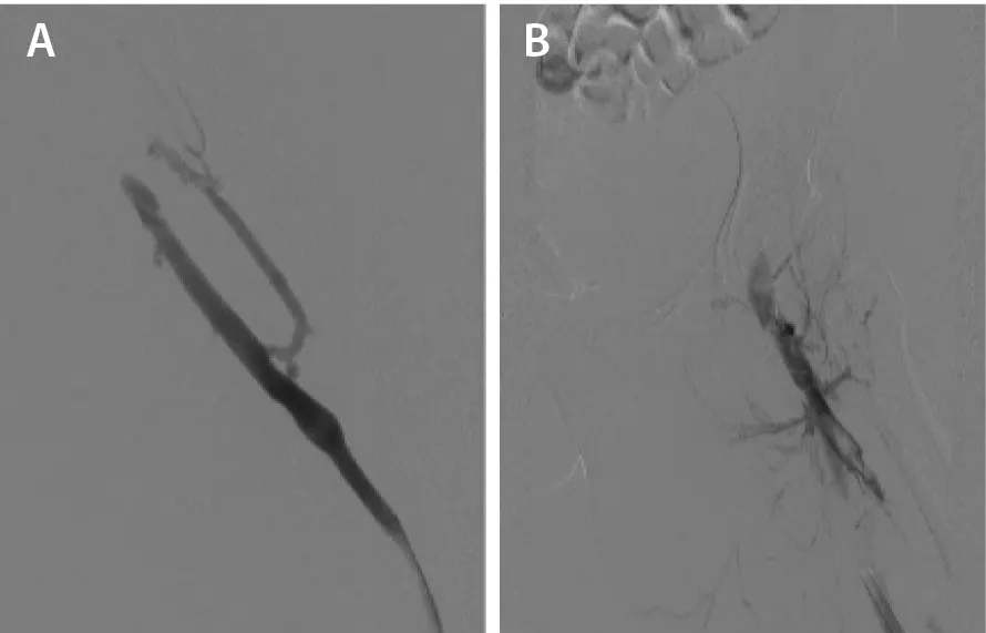
Figure 1. Left popliteal vein (A) and left FV pretreatment (B).
Device Selection and Settings
A 12-Fr Liberant™ thrombectomy system catheter (Medtronic) was selected to remove the thrombus in the left iliofemoral vein. Three passes were advanced using the Liberant device. Standard mode was used for the first two passes. Boost mode was used for the last pass in the left common iliac vein (CIV) given the large vein size and anticipated large degree of clot burden.
Thrombectomy Technique
The catheter was advanced while rotating the device to utilize the angled tip to aid in mural thrombus removal. Thrombectomy was largely performed without the use of a wire and the Liberant catheter tracked without resistance through the entire iliofemoral segment. A 0.035-inch wire was utilized to cross into the IVC at the origin of the left CIV due to severe compression.
Repeated venograms demonstrated an area of adherent thrombus in the left FV. Excipio™* LV Prime thrombectomy device (Contego Medical) was utilized to remove this adherent thrombus. The basket was opened proximally to the area of thrombus and then pulled back while aspiration was active in the Liberant catheter. This was repeated twice for a successful thrombus extraction.
Post-Thrombectomy Treatment
An Amplatz stiff guidewire was exchanged into the IVC. Intravascular ultrasound (IVUS) was performed on the treated segment, which demonstrated resolution of all significant thrombus burden. IVUS demonstrated multiple areas of left iliac vein compression and severe narrowing at the origin of the left CIV (Figure 2A). A 16- X 150-mm Abre™ venous self-expanding stent system (Medtronic) was deployed into the left CIV and left external iliac vein and postdilated with a 16-mm balloon. Repeat IVUS demonstrated resolution of the left CIV compression (Figure 2B). Final venography demonstrated resolution of all stenoses with brisk flow into the IVC (Figure 3). Total blood loss for the procedure was under 250 mL.
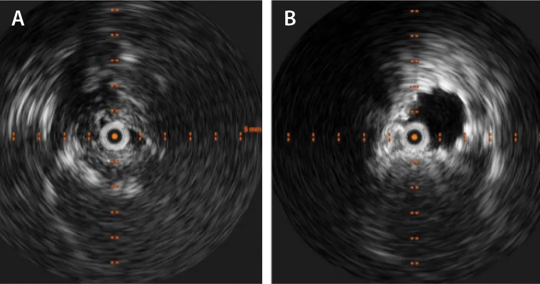
Figure 2. IVUS of the left CIV before (A) and after (B) stenting.
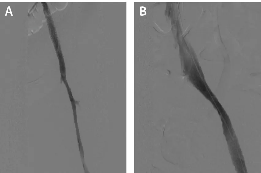
Figure 3. Left FV (A) and left iliac vein (B) posttreatment.
Postprocedure Course
The patient did well postoperatively. She was maintained on IV heparin on postoperative day 0. Her swelling and pain significantly improved. She was discharged on postoperative day 1 with apixaban and compression stockings. She was most recently seen at her 6-month clinic follow-up with complete symptom resolution. An in-office venous DUS at 6-month follow-up showed a patent left iliac vein stent and iliofemoral venous system.
REFLECTIONS
The Liberant thrombectomy system met my expectations of safety and performance in removal of thrombus in the lower extremity. In our experience, the device has been efficient at removing large thrombus burden in both the arterial and venous system with an acceptably low amount of blood loss. It has been our preference to use the device in standard mode with selective use of boost mode in areas of large thrombus burden. We use Excipio LV Prime as a mechanical adjunct to facilitate removal of adherent and mural thrombus when using the Liberant system.
Disclosures: Consultant to Medtronic and Penumbra, Inc.
Advertisement
Advertisement