Advertisement
Advertisement
October 2026
Combined Transarterial Embolization and Percutaneous Ablation for Renal Cell Carcinoma
Patient selection, embolization technique, procedural workflow, and clinical outcomes with preablation TAE for large, hypervascular, and anatomically complex RCC.
By Francis G. Celii, MD; Justin A. Ahrar; and Mohamed E. Abdelsalam, MD
Percutaneous image-guided thermal ablation is an established nephron-sparing treatment for patients with cT1a renal cell carcinoma (RCC).1-3 Percutaneous cryoablation, microwave ablation (MWA), and radiofrequency ablation (RFA) provide excellent oncologic outcomes in appropriately selected patients.4-12 When compared with partial nephrectomy, image-guided ablation has the added advantage of preserving renal function and reducing procedural morbidity.13,14
While technical success and local tumor control rates are often reported to exceed 90% for cT1a RCC, treatment efficacy declines as tumor size and complexity increase.4,8,15,16 Tumors > 3 cm, particularly cT1b and cT2a lesions, often require multiple overlapping ablations and are associated with higher rates of residual disease, local recurrence, and hemorrhage.15,16 Hypervascular clear cell RCC further complicates thermal ablation because tumor perfusion reduces energy deposition and increases bleeding risk, especially in central or endophytic tumors.17,18
Selective transarterial embolization (TAE) has emerged as an adjunct to overcome these limitations. By reducing tumor vascularity before ablation, embolization may decrease risk of hemorrhage, improve visualization of endophytic lesions, minimize heat-sink effect, and facilitate more complete treatment of larger or anatomically complex tumors.19-27
This article summarizes current evidence regarding patient selection, embolization techniques, embolic materials, and clinical outcomes of combined TAE and percutaneous ablation for RCC.
PATIENT SELECTION
Combined TAE and percutaneous ablation is best suited for patients with larger, hypervascular, or anatomically complex renal tumors in whom ablation alone may be technically challenging or associated with a higher risk of complications.22,24,26
Tumor size is the primary determinant for the selection of this combination therapy (Figure 1). Percutaneous ablation alone provides excellent local control for most exophytic cT1a renal tumors < 3 cm. As tumor size increases, particularly in cT1b lesions (4-7 cm), complete ablation becomes more challenging, with lower primary technical efficacy and higher rates of local tumor progression.15,28 Consequently, most published studies of combined TAE and ablation have focused on tumors measuring ≥ 3 cm and cT1b lesions.20,22-24,26,27
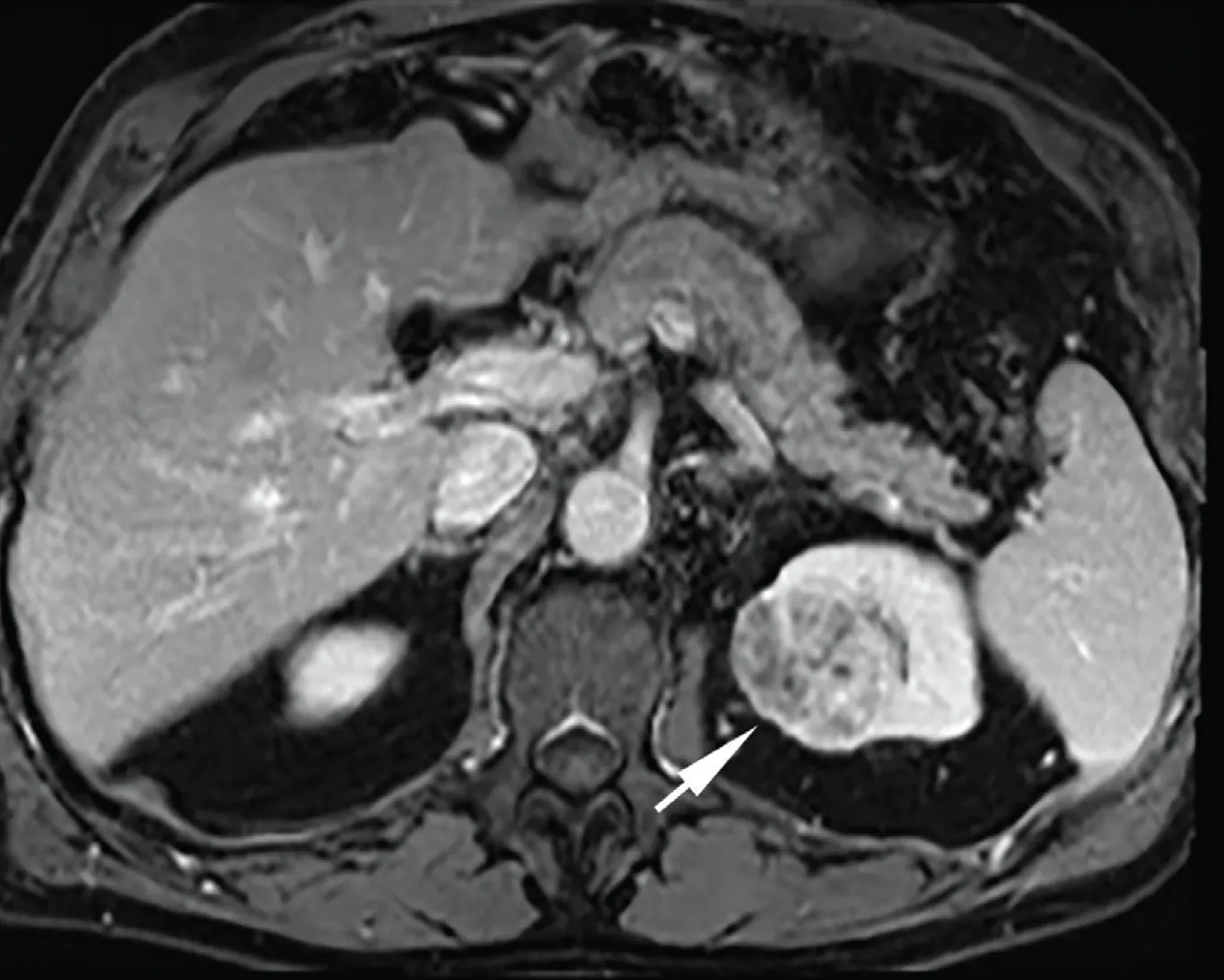
Figure 1. Axial contrast-enhanced T1-weighted MRI of the abdomen in a man in his early 80s demonstrated a 4.7-cm solid enhancing mass (arrow) in the medial upper pole of the left kidney. Biopsy demonstrated clear cell RCC, grade 2. Given the tumor’s large size and hypervascularity, the patient underwent TAE followed by cryoablation during a single treatment session.
Tumor location and vascularity further influence patient selection. Endophytic and central tumors are more difficult to target because of limited CT conspicuity, and they are more challenging to ablate considering their proximity to larger renal vessels.15 Embolization improves lesion visualization, reduces tumor perfusion, and may lessen heat-sink effect during thermal ablation.21-23 Hypervascular clear cell RCC also carries a greater risk of hemorrhage during placement of multiple applicators, providing an additional rationale for preablation embolization.29
Based on the available evidence, adjunctive embolization may be considered for cT1b, hypervascular, endophytic, or centrally located tumors, whereas most small exophytic cT1a lesions can be treated successfully with ablation alone.30 In our practice, we consider TAE for tumors > 5 cm, as well as for endophytic and hypervascular tumors.
EMBOLIZATION TECHNIQUE AND EMBOLIC MATERIALS
Successful combined therapy depends on superselective embolization that devascularizes the target tumor while preserving uninvolved renal parenchyma (Figure 2A-2C). Embolization is performed using femoral or radial arterial access immediately before or within several days prior to ablation. Selective angiography of the renal artery helps localize tumor-feeding vessels (Figure 2A). These branches are then catheterized using a standard microcatheter and microwire technique, allowing for superselective embolization of the tumor (Figure 2B). The endpoint for embolization is elimination of the tumor blush or near-complete stasis while maintaining perfusion to the remaining normal kidney (Figure 2C).23-25
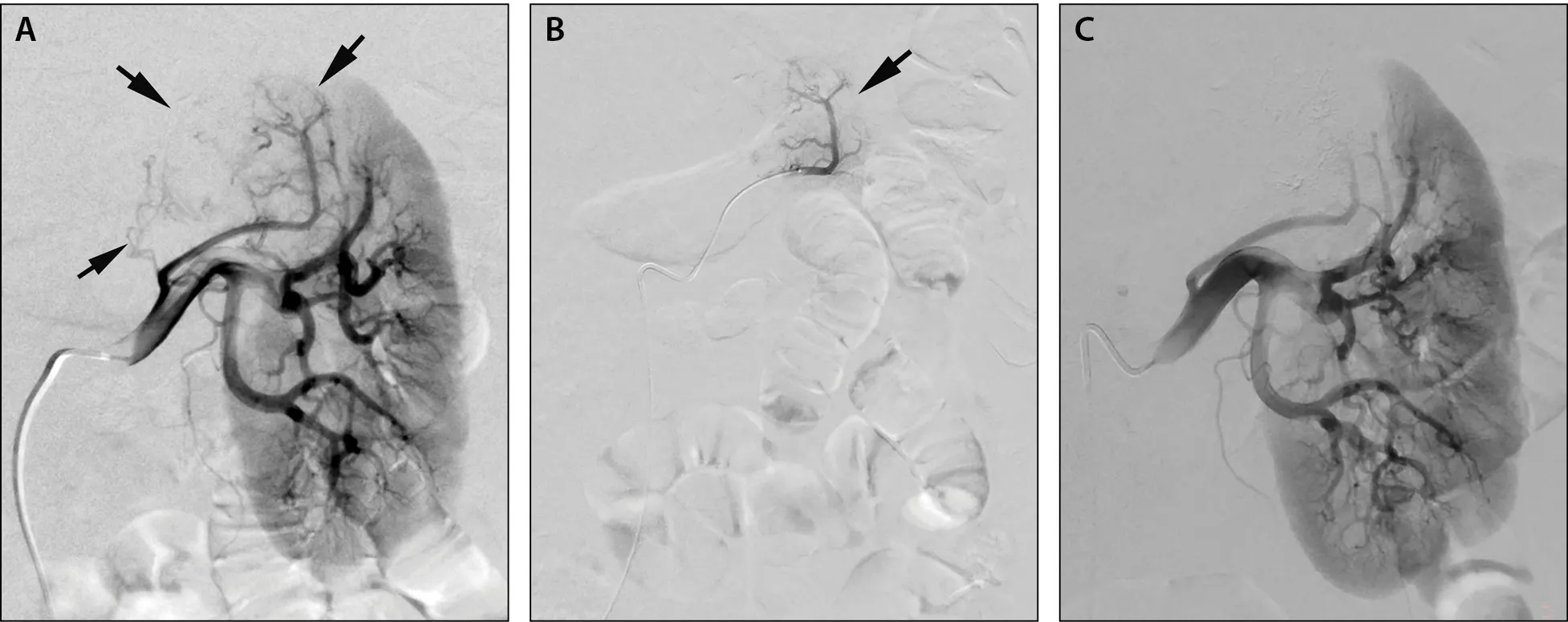
Figure 2. Selective angiography of the left renal artery shows neovascularity and tumor blush (arrows) in the upper pole of the left kidney (A). Superselective angiography of the largest feeding artery shows tumor blush (arrow); embolization of this artery and two smaller, more medial feeding arteries (not shown) was carried out using 40- and 400-μm microsphere particles (B). Selective angiography of the left kidney after embolization demonstrates complete elimination of the neovascularity and tumor blush in the upper pole of the left kidney, with preservation of perfusion to the remainder of the kidney (C).
A variety of embolic agents have been used, including ethanol with or without iodized oil, gelatin sponge, polyvinyl alcohol (PVA) particles, N-butyl cyanoacrylate glue, and microspheres.19,20,24,27 Ethanol provides permanent microvascular occlusion, whereas gelatin sponge offers temporary embolization. Terauchi et al compared a combination of ethanol and iodized oil to a regimen of gelatin sponge followed by iodized oil.25 There were no differences in the clinical outcomes for TAE, but patients in the ethanol group reported a significantly higher incidence of pain during the procedure (P < .05). PVA particles have also been used successfully before cryoablation of large tumors. Iodized oil may be used alone or together with other embolic agents to mark the tumor, improving CT visualization of endophytic lesions at the time of ablation.19,23 However, residual high-density iodized oil remains within the tumor and may complicate interpretation of follow-up CT imaging studies after ablation. Microspheres may also be visualized within the tumor on CT images shortly after embolization (Figure 3A), but they do not interfere with or complicate subsequent follow-up imaging. Based on published reports, the choice of embolic agent largely depends on operator preference and the availability of embolic products.
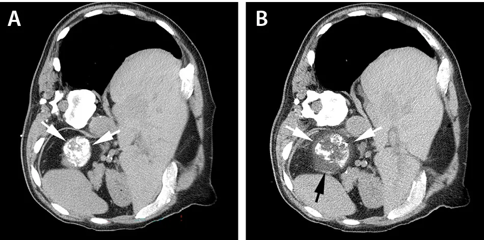
Figure 3. Immediately after embolization, the patient was placed in the left lateral decubitus position. Axial noncontrast CT demonstrates retention of contrast and embolic material within the tumor (arrows), facilitating precise probe placement under CT guidance (A). Cryoablation was performed using six cryoprobes; axial noncontrast CT demonstrates a low-attenuation iceball encompassing the entire tumor with adequate ablation margin (arrows) (B).
The optimal interval between embolization and ablation has not yet been clearly established. Most published series report ablation during the same hospitalization, typically within several hours to several days after embolization. However, the reported interval varies considerably, ranging from a few hours to 46 days.22,24,31 Many institutions perform embolization and ablation in separate angiography and CT suites, making same-session treatment logistically challenging. At our institution, a hybrid interventional suite equipped with angiography and CT-on-rails allows both procedures to be performed during a single patient encounter. To expedite the procedure further, we access the left radial artery with the patient in the prone position, eliminating the need for patient repositioning before CT-guided ablation. When the patient remains in the same position for both procedures, room time is significantly shorter.32 Completing both TAE and ablation in one session precludes the need for two separate encounters and anesthesia events. It is a patient-centric practice and likely improves patient experience.
PERCUTANEOUS ABLATION TECHNIQUE
The choice of ablative modality often depends on tumor size, location, and adjacent anatomy.33 The availability of devices and operator experience also have a large impact on the choice of ablation technology. Cryoablation, MWA, and RFA have all been combined successfully with TAE.19,23,24,30,34 Cryoablation offers the additional advantage of real-time visualization of the treatment zone, as the expanding iceball is readily visible on CT imaging (Figure 3B). Regardless of the energy source, the objective is complete tumor destruction with an ablative margin ≥ 5 mm while preserving normal renal parenchyma and kidney function (Figure 4).1-4
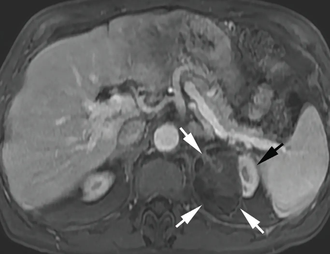
Figure 4. Axial contrast-enhanced T1-weighted MRI of the abdomen obtained 6 weeks after ablation demonstrates complete absence of enhancement within the ablation zone (white arrows). The lateral aspect of the upper pole of the left kidney is preserved and demonstrates normal enhancement (black arrow).
Embolization does not fundamentally alter ablation technique but facilitates treatment of larger, more complex tumors by reducing vascularity, improving lesion conspicuity, and decreasing hemorrhagic risk.22-24 Successful combined therapy therefore depends on integrating meticulous embolization with established principles of image-guided renal tumor ablation.
CLINICAL OUTCOMES
Published experience with combined TAE and percutaneous ablation for RCC is limited to retrospective single-center series, pilot studies, and one prospective cohort study (Table 1).6,19-27,31,34 Despite these limitations, available evidence suggests that combined therapy is technically feasible and provides favorable local tumor control, while preserving renal function. The combined treatment approach also has an acceptable safety profile in carefully selected patients with larger, hypervascular, or anatomically complex tumors.6,19-27,31
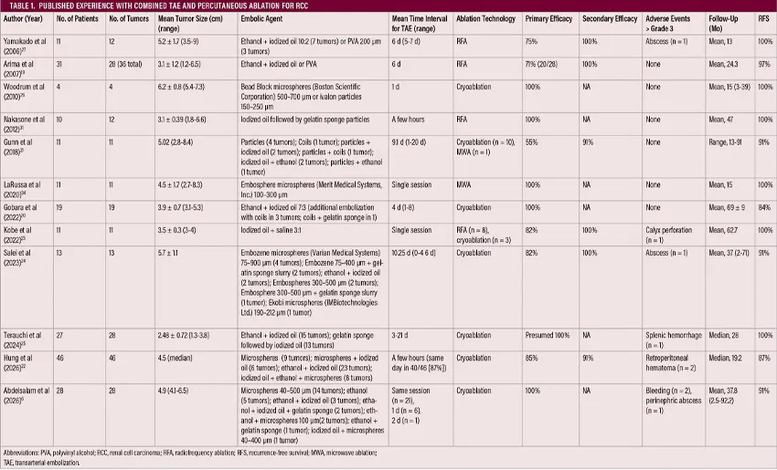
Technical success for the combined therapy has been consistently high across published studies. Nakasone et al reported complete RFA without residual enhancement in all treated tumors, while Gobara et al achieved complete technical success with embolization followed by cryoablation for tumors ≥ 3 cm.20,31 Abdelsalam et al reported 100% primary efficacy in patients with T1b tumors.6 Long-term outcomes have also been encouraging, with Gobara et al reporting only two local recurrences during a median follow-up of 68 months, no distant metastases, and a 5-year cancer-specific survival of 100%.20 Similarly, Salei et al observed > 90% recurrence-free survival following embolization and cryoablation of T1b and T2a tumors.24
A major benefit of adjunctive embolization is reduced hemorrhage during and immediately after ablation. Woodrum et al studied hematoma volume after cryoablation for tumors > 5 cm with or without preablation TAE, demonstrating significant reduction in postprocedural hematoma volume in patients treated with preablation TAE.26 Salei et al compared a combined TAE and cryoablation approach to cryoablation alone in patients with T1b and T2a tumors.24 Patients in the combined group had significantly larger tumors (P = .0003) with higher nephrometry scores (P = .0192). Despite these unfavorable tumor characteristics, the combined group had lower rates of major hemorrhagic complications.
Across published studies, superselective embolization combined with nephron-sparing ablation has resulted in little or no deterioration of renal function, supporting preservation of functional renal parenchyma.20,23,24,26,31 Major complications are uncommon when careful patient selection, meticulous procedural planning, and superselective embolization are employed. Nontarget embolization is rare and can be minimized through detailed angiographic evaluation, precise microcatheter positioning, and controlled embolic delivery.19-27
CONCLUSION
Percutaneous ablation offers an effective nephron-sparing approach for cT1a RCC; however, larger and hypervascular tumors, particularly those in anatomically challenging locations, can be more difficult to treat completely. Preablation TAE provides a complementary strategy that reduces tumor perfusion and vascularity, potentially improving ablation efficacy while reducing hemorrhagic risk.
1. Campbell SC, Clark PE, Chang SS, et al. Renal mass and localized renal cancer: evaluation, management, and follow-up: AUA guideline: part I. J Urol. 2021;206:199-208. doi: 10.1097/JU.0000000000001911
2. Finelli A, Ismaila N, Bro B, et al. Management of small renal masses: American Society of Clinical Oncology clinical practice guideline. J Clin Oncol. 2017;35:668-680. doi: 10.1200/JCO.2016.69.9645
3. Motzer RJ, Jonasch E, Agarwal N, et al. NCCN Guidelines® insights: kidney cancer, version 2.2024. J Natl Compr Canc Netw. 2024;22:4-16. doi: 10.6004/jnccn.2024.0008
4. Aarts BM, Prevoo W, Meier MAJ, et al. Percutaneous microwave ablation of histologically proven T1 renal cell carcinoma. Cardiovasc Intervent Radiol. 2020;43:1025-1033. doi: 10.1007/s00270-020-02423-7
5. Abdelsalam ME, Awad A, Baiomy A, et al. Outcomes of radiofrequency ablation for solitary T1a renal cell carcinoma: a 20-year tertiary cancer center experience. Cancers (Basel). 2023;15:909. doi: 10.3390/cancers15030909
6. Abdelsalam ME, Awad A, Kang F, et al. Percutaneous cryoablation of clinical T1b renal tumors: a tertiary cancer center experience. Cardiovasc Intervent Radiol. Published online March 17, 2026. doi: 10.1007/s00270-026-04381-y
7. Abdelsalam ME, Mecci N, Awad A, et al. Magnetic-resonance-imaging-guided cryoablation for solitary-biopsy-proven renal cell carcinoma: a tertiary cancer center experience. Cancers (Basel). 2024;16:1815. doi: 10.3390/cancers16101815
8. Atwell TD, Vlaminck JJ, Boorjian SA, et al. Percutaneous cryoablation of stage T1b renal cell carcinoma: technique considerations, safety, and local tumor control. J Vasc Interv Radiol. 2015;26:792-799. doi: 10.1016/j.jvir.2015.02.010
9. Breen DJ, King AJ, Patel N, et al. Image-guided cryoablation for sporadic renal cell carcinoma: three- and 5-year outcomes in 220 patients with biopsy-proven renal cell carcinoma. Radiology. 2018;289:554-561. doi: 10.1148/radiol.2018180249
10. Guo J, Arellano RS. Percutaneous microwave ablation of category T1a renal cell carcinoma: intermediate results on safety, technical feasibility, and clinical outcomes of 119 tumors. AJR Am J Roentgenol. 2021;216:117-124. doi: 10.2214/AJR.20.22818
11. Marshall HR, Shakeri S, Hosseiny M, et al. Long-term survival after percutaneous radiofrequency ablation of pathologically proven renal cell carcinoma in 100 patients. J Vasc Interv Radiol. 2020;31:15-24. doi: 10.1016/j.jvir.2019.09.011
12. Yu J, Wang H, Cheng ZG, et al. A multicenter 10-year oncologic outcome of ultrasound-guided percutaneous microwave ablation of clinical T1 renal cell carcinoma: will it stand the test of time? Eur Radiol. 2022;32:89-100. doi: 10.1007/s00330-021-07900-2
13. Mason RJ, Atwell TD, Lohse C, et al. Renal functional outcomes in patients undergoing percutaneous cryoablation or partial nephrectomy for a solitary renal mass. BJU Int. 2017;120:544-549. doi: 10.1111/bju.13917
14. Pierorazio PM, Johnson MH, Patel HD, et al. Management of renal masses and localized renal cancer: systematic review and meta-analysis. J Urol. 2016;196:989-999. doi: 10.1016/j.juro.2016.04.081
15. King AJ, Steenkamp R, Graumann O, et al. Risk factors for residual unablated tumour following CT-guided percutaneous renal cryoablation: lessons from the EuRECA registry. Cardiovasc Intervent Radiol. 2025;48:196-204. doi: 10.1007/s00270-024-03951-2
16. Mouli SK, McDevitt JL, Su YK, et al. Analysis of the RENAL and mRENAL scores and the relative importance of their components in the prediction of complications and local progression after percutaneous renal cryoablation. J Vasc Interv Radiol. 2017;28:860-867. doi: 10.1016/j.jvir.2016.12.1224
17. Haddad MM, Schmit GD, Kurup AN, et al. Percutaneous cryoablation of solitary, sporadic renal cell carcinoma: outcome analysis based on clear-cell versus papillary subtypes. J Vasc Interv Radiol. 2018;29:1122-1126. doi: 10.1016/j.jvir.2018.02.029
18. Lay AH, Faddegon S, Olweny EO, et al. Oncologic efficacy of radio frequency ablation for small renal masses: clear cell vs papillary subtype. J Urol. 2015;194:653-657. doi: 10.1016/j.juro.2015.03.115
19. Arima K, Yamakado K, Kinbara H, et al. Percutaneous radiofrequency ablation with transarterial embolization is useful for treatment of stage 1 renal cell carcinoma with surgical risk: results at 2-year mean follow up. Int J Urol. 2007;14:585-590; discussion 90. doi: 10.1111/j.1442-2042.2007.01740.x
20. Gobara H, Matsui Y, Uka M, et al. Percutaneous cryoablation combined with prior transcatheter arterial embolization for renal cell carcinomas of 3 cm or larger: a prospective study. Int J Clin Oncol. 2022;27:1589-1595. doi: 10.1007/s10147-022-02224-7
21. Gunn AJ, Mullenbach BJ, Poundstone MM, et al. Trans-arterial embolization of renal cell carcinoma prior to percutaneous ablation: technical aspects, institutional experience, and brief review of the literature. Curr Urol. 2018;12:43-49. doi: 10.1159/000447230
22. Hung ML, Wang R, Yu C, et al. Efficacy and safety of sequential transarterial embolization and cryoablation of renal masses greater than 3 cm. Urol Oncol. 2026;44:110964. doi: 10.1016/j.urolonc.2025.11.018
23. Kobe A, Tselikas L, Deschamps F, et al. Thermal ablation combined with selective transarterial embolization of centrally located renal cell carcinomas measuring 3 cm or larger. Cardiovasc Intervent Radiol. 2022;45:371-379. doi: 10.1007/s00270-021-03036-4
24. Salei A, Raymond M, Savage C, et al. Transarterial embolization of T1b and T2a renal cell carcinoma prior to percutaneous cryoablation: a retrospective comparative study. Abdom Radiol (NY). 2023;48:773-779. doi: 10.1007/s00261-022-03755-3
25. Terauchi M, Yamashiro T, Sawamura S, et al. Combining transarterial embolization and percutaneous cryoablation for early-stage renal cell carcinoma: embolization materials and impacts of tumor size. Tomography. 2024;10:1767-1779. doi: 10.3390/tomography10110130
26. Woodrum DA, Atwell TD, Farrell MA, et al. Role of intraarterial embolization before cryoablation of large renal tumors: a pilot study. J Vasc Interv Radiol. 2010;21:930-936. doi: 10.1016/j.jvir.2010.02.015
27. Yamakado K, Nakatsuka A, Kobayashi S, et al. Radiofrequency ablation combined with renal arterial embolization for the treatment of unresectable renal cell carcinoma larger than 3.5 cm: initial experience. Cardiovasc Intervent Radiol. 2006;29:389-94. doi: 10.1007/s00270-004-0090-9
28. Garnon J, Van Strijen MJ, Nielsen TK, et al. Safety of percutaneous renal cryoablation: an international multicentre experience from the EuRECA retrospective percutaneous database. Eur Radiol. 2019;29:6293-6399. doi: 10.1007/s00330-019-06191-y
29. Okhunov Z, Moreira DM, Del Junco M, et al. Predictors of complications after percutaneous image-guided renal cryoablation for T1a renal cortical neoplasms. J Endourol. 2017;31:7-13. doi: 10.1089/end.2016.0684
30. Gunn AJ, Parikh NS, Bhatia S. Society of Interventional Radiology quality improvement standards on percutaneous ablation in renal cell carcinoma. J Vasc Interv Radiol. 2020;31:195-201.e3. doi: 10.1016/j.jvir.2019.11.004
31. Nakasone Y, Kawanaka K, Ikeda O, et al. Sequential combination treatment (arterial embolization and percutaneous radiofrequency ablation) of inoperable renal cell carcinoma: single-center pilot study. Acta Radiol. 2012;53:410-414. doi: 10.1258/ar.2012.110413
32. Celii FG, Abdelsalam ME, Foss WT, et al. Safety and efficacy of single-session, single-position (prone) combined trans-radial renal embolization and ablation treatment of cT1 renal cell carcinoma. Presented at: GEST 2024; May 16-19, 2024; New York, New York.
33. Abdelsalam ME, Ahrar K, Sheth RA, et al. Interventional oncology: a primer for clinicians on the role of ablation and embolization for solid tumors. CA Cancer J Clin. 2026;76:e70051. doi: 10.3322/caac.70051
34. LaRussa S, Vanden Berg RW, Craig KM, et al. Is there a role for combination, single-session selective transarterial embolization and microwave ablation for large renal masses? Cardiovasc Intervent Radiol. 2020;43:1468-1473. doi: 10.1007/s00270-020-02568-5
Advertisement
Advertisement