Advertisement
Advertisement
September 2026
Sponsored by Endologix
Optimizing Patient Selection for the DETOUR™ System
Dr. Amit Dwivedi offers his perspective on patient selection criteria for the DETOUR System, including patient and lesion characteristics, key lessons learned, common misconceptions, and advice to physicians prior to their first use of the device, and shares a case example highlighting use of the DETOUR System.

Has your patient selection evolved since your first case with the DETOUR™ System (Endologix)?
Yes, my approach to patient selection has evolved since I first started using the DETOUR System. I adopted the technology after my first case treating a morbidly obese patient with uncontrolled diabetes, claudication pain (walk distance, a half a block), and an ankle-brachial index (ABI) of 0.54 at presentation. Postprocedure, the claudication pain had resolved and ABI improved to 0.9, which has been maintained over 3-year follow-up. Subsequently, I utilized the DETOUR System on the patient described in the case study (see Case Study Sidebar), as well as five additional cases who met my patient selection criteria, as I describe below. All of these patients have completed at least 2 years of follow-up and have done well.
What patient or lesion characteristics make you think of DETOUR immediately?
Characteristics I look for in patients include those who are frail, heavy smokers, immunosuppressed, morbidly obese, or on certain medications like steroids, as well as those who have cancer, poorly controlled diabetes, a hostile groin, or burns and scars.
For lesions, I consider the DETOUR System in patients with TransAtlantic Inter Society Consensus (TASC) D lesions within the superficial femoral artery (SFA) and in cases of redo stenting.*
When do you choose DETOUR instead of bypass surgery?
The previously noted patient and lesion characteristics typically guide my choice to employ the DETOUR System rather than bypass surgery.
What have your biggest patient selection lessons been over time?
Some lessons I’ve learned include:
- Select patients with a minimum 1-cm SFA stump
- Avoid extremely calcified areas for entry from artery to vein
- Avoid patients with deep vein thrombosis (DVT) or prior DVT
- Avoid single-vessel outflow at the tibial vessels as an initial case
- Perform follow-up duplex ultrasound before discharge
What misconceptions do physicians have about the ideal DETOUR patient?
The biggest misconceptions include the risk of DVT by stenting in superficial femoral vein (SFV), bypass surgery options being hindered because of DETOUR, and that the procedure can be technically challenging. It should be noted that any new technology has to be given fair trial and time to be understood and mastered.
Have you moved DETOUR earlier in your treatment algorithm? Why?
Yes, I consider using DETOUR earlier in my treatment algorithm based on lesion and patient characteristics, as the patient outcomes seem to improve.
What advice would you give a physician selecting their first DETOUR patient?
Start with patients with a proximal SFA that is open at least 8 cm with good contralateral access and minimal aortoiliac occlusive disease. Ensure there is no calcification at the entry points, consider the orientation of artery to vein, and ensure there is an adequate landing zone as well as appropriate two- to three-vessel runoff. Postoperatively, ensure that the patient is on antiplatelet therapy with a statin and anticoagulation and have a plan for appropriate follow-up with duplex ultrasound and ABI measurements.
In your experience using the DETOUR System, how important is it for the broader care team—including nurses, techs, and ultrasound staff—to be educated on appropriate patient selection for newer therapies?
The techs, nurses, and ultrasound staff are an important part of the team, so educating them about the device, their role during cases, and clinical evaluation postoperatively—specifically to alert physicians of any untoward outcomes—is crucial to overall success.
CASE STUDY: USE OF THE DETOUR SYSTEM IN A PATIENT WITH PRIOR FEMORAL ENDARTERECTOMY AND LEFT SFA OCCLUSION
CASE PRESENTATION
A female patient in her mid-60s with a previous left femoral endarterectomy presented with claudication pain in the left calf while walking. She had a history of coronary artery disease and hypertension and was a 1 pack/day smoker but had stopped smoking for 6 months prior to presentation. Claudication distance was three-quarters of a block and her ABI was 0.6 in the left leg and 0.97 in the right leg. CTA showed a left SFA occlusion with a dense scar in the left groin (Figure 1).
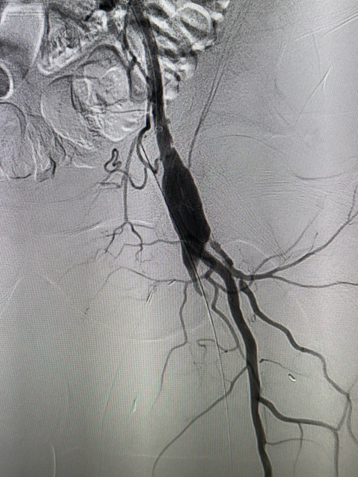
Figure 1. Occluded left SFA with previous patch repair of the CFA.
PROCEDURAL OVERVIEW
The right common femoral artery (CFA) was accessed with an 8-F sheath, and an aortogram was obtained. An Omni Flush catheter (AngioDynamics, Inc.) was used to gain access to the left external iliac artery, and the short 8-F sheath was then exchanged for an 8-F, 45-cm sheath. Selective angiography of the left lower extremity was performed, and using an angled catheter and 0.014-inch wire, access was gained to the left SFA.
The first 4 cm of the SFA was ballooned with a 5-mm X 4-cm low-pressure balloon. The ENDOCROSS™ Device (Endologix) was then introduced, and with appropriate orientation, the needle was fired to gain access in the left SFV at the level of the lesser trochanter. Access was obtained in the left posterior tibial vein under ultrasound guidance and a 6-F, 25-cm sheath was placed. A snare was introduced through the 6-F sheath and placed at level of the lesser trochanter. The wire from the ENDOCROSS Device was then snared and brought out of the 6-F sheath. Over this through-and-through wire, a 4-mm X 4-cm balloon was introduced at the entry site from artery into vein and the opening was dilated. The ENDOCROSS Device was then reintroduced over the through-and-through wire into the femoral vein and, at 2 cm above thepatella, the device was refired with appropriate orientation to gain access into the popliteal vein (Figure 2). The ENDOCROSS Device was removed, and the distal entry point was dilated with a 4-mm X 4-cm balloon. The 0.014-inch wire was exchanged over a crossing catheter to a 0.035-inch wire. The through-and-through wire was removed, and TORUS™ Stent Grafts (Endologix) were placed all the way from the origin of the SFA to the popliteal artery with adequate overlap (Figure 3, Figure 4, and Figure 5), and the overlaps and proximal and distal ends of the stents were ballooned. Completion angiography confirmed procedural success and the sheaths were removed.
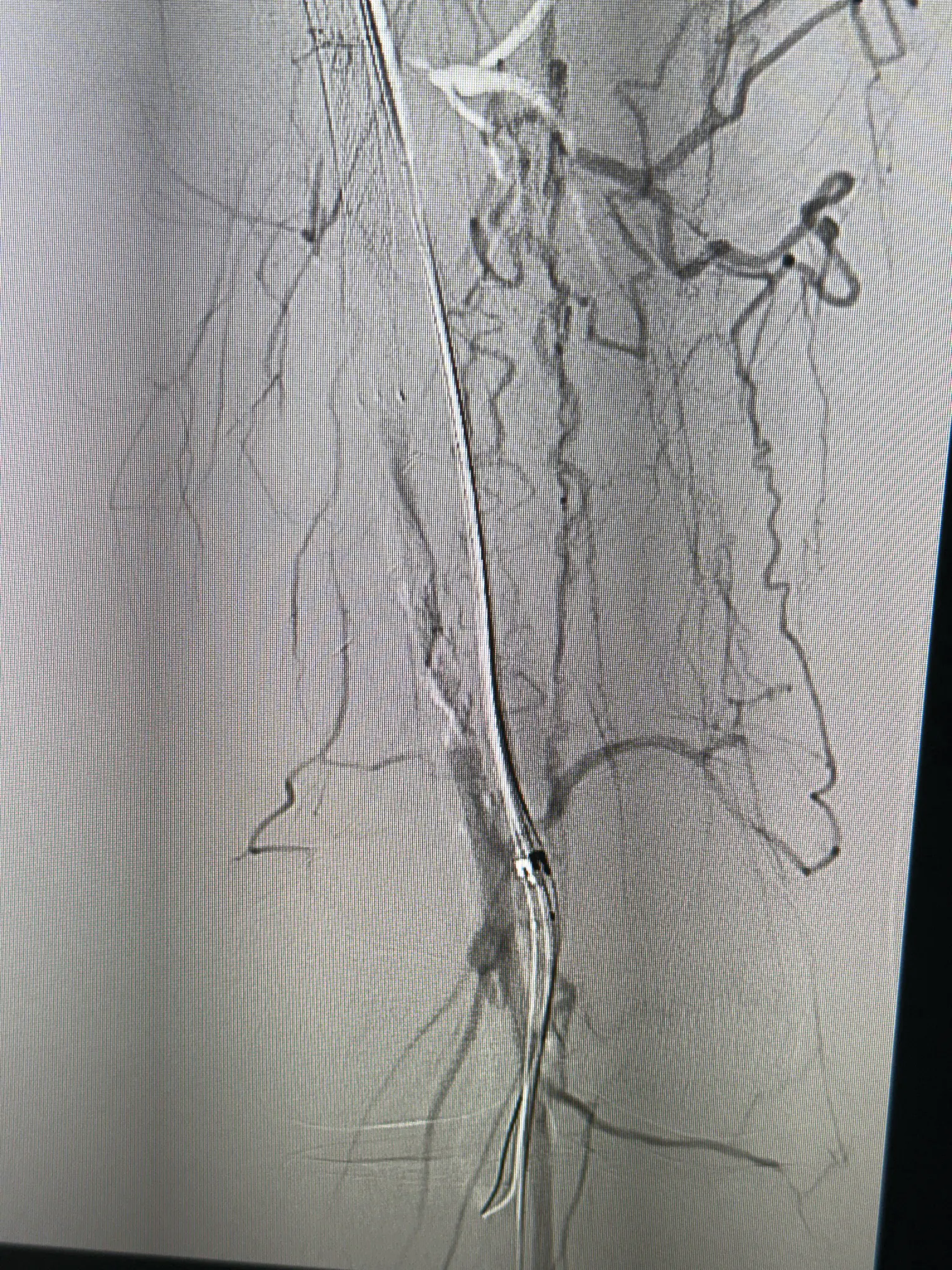
Figure 2. DETOUR System crossing from the popliteal vein to popliteal artery at the level above the patella.
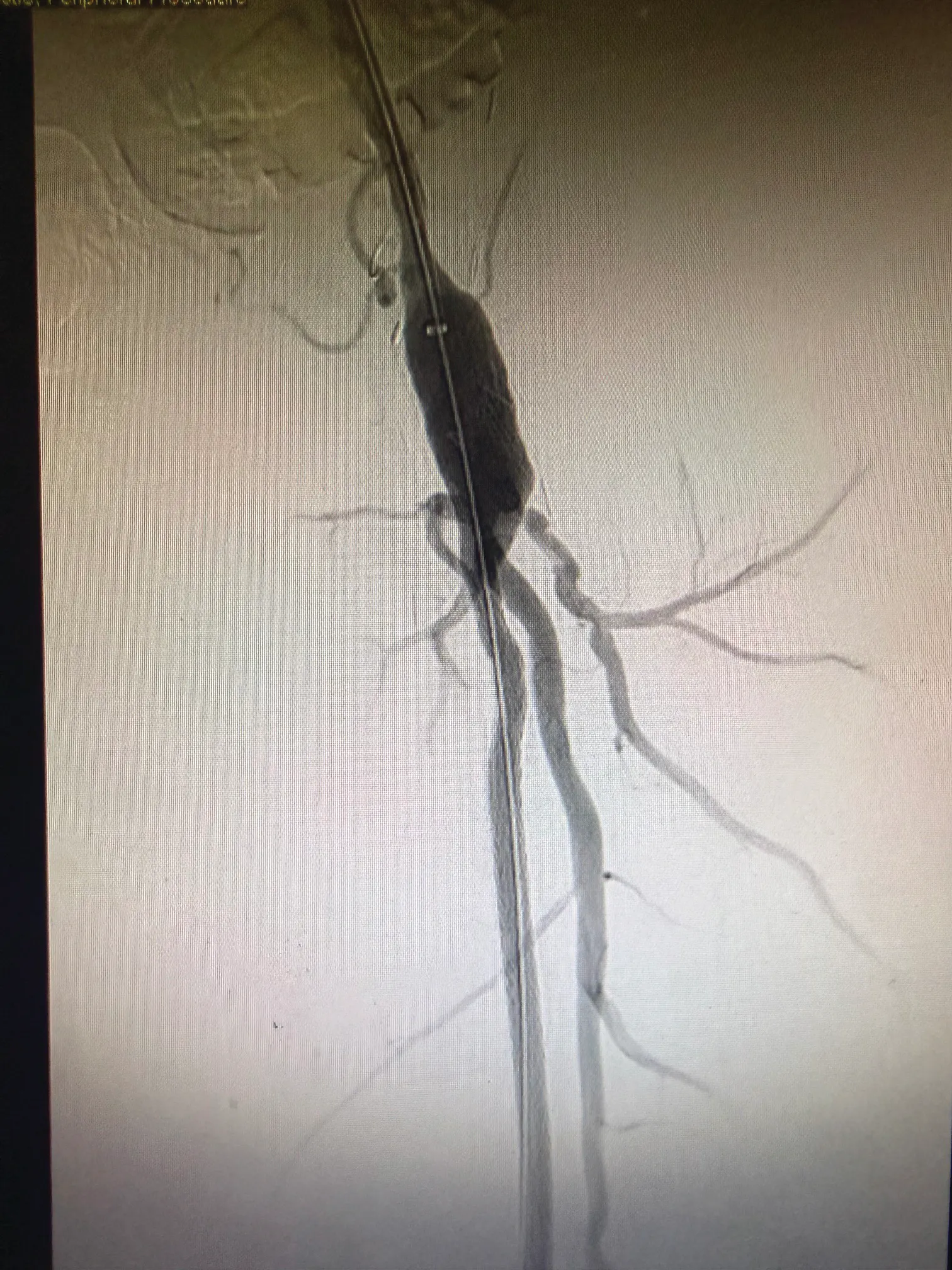
Figure 3. Placement of the TORUS Stent Graft at the origin for the left SFA.
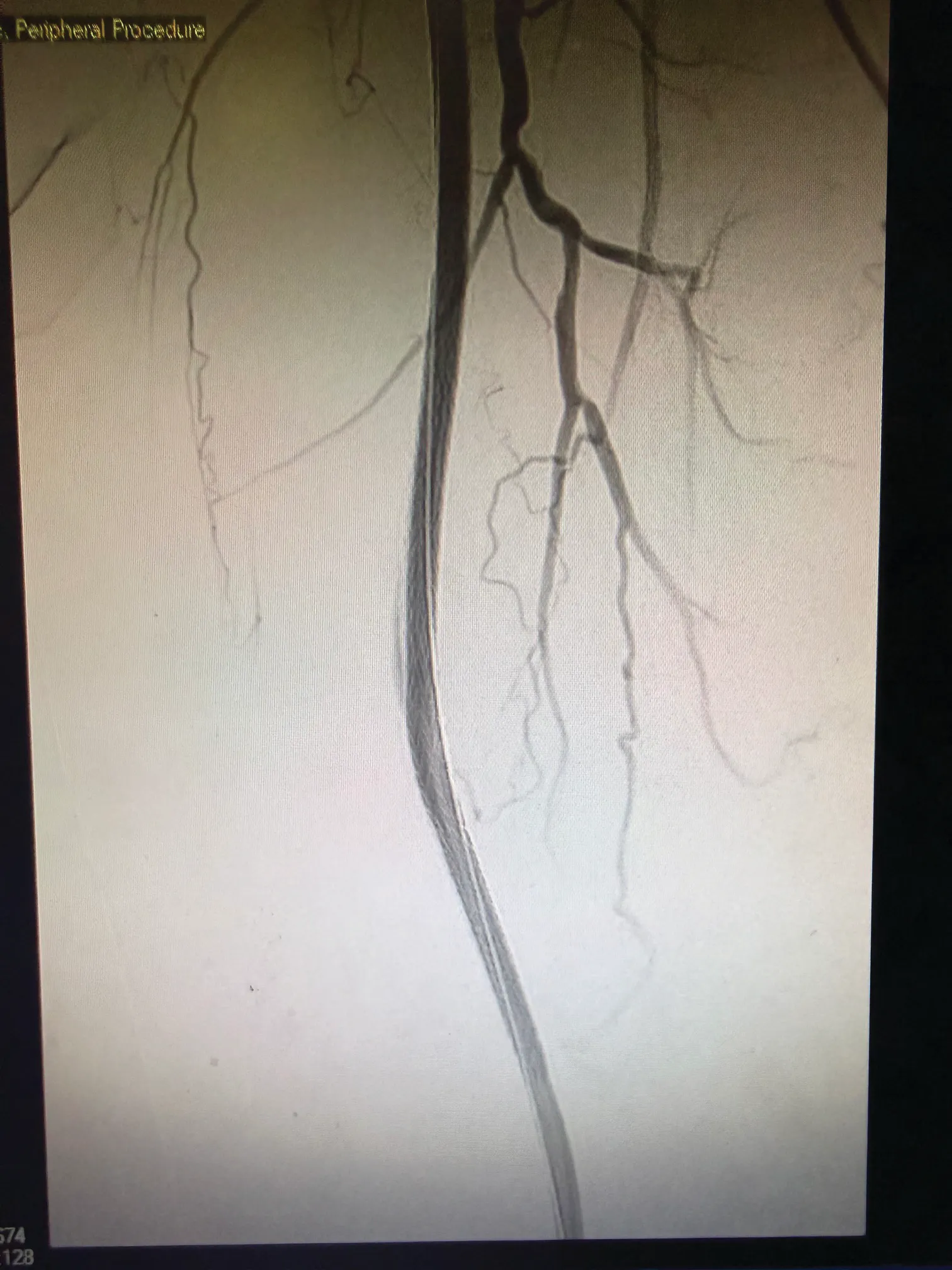
Figure 4. TORUS Stent Graft in the left SFV in the mid to lower thigh.
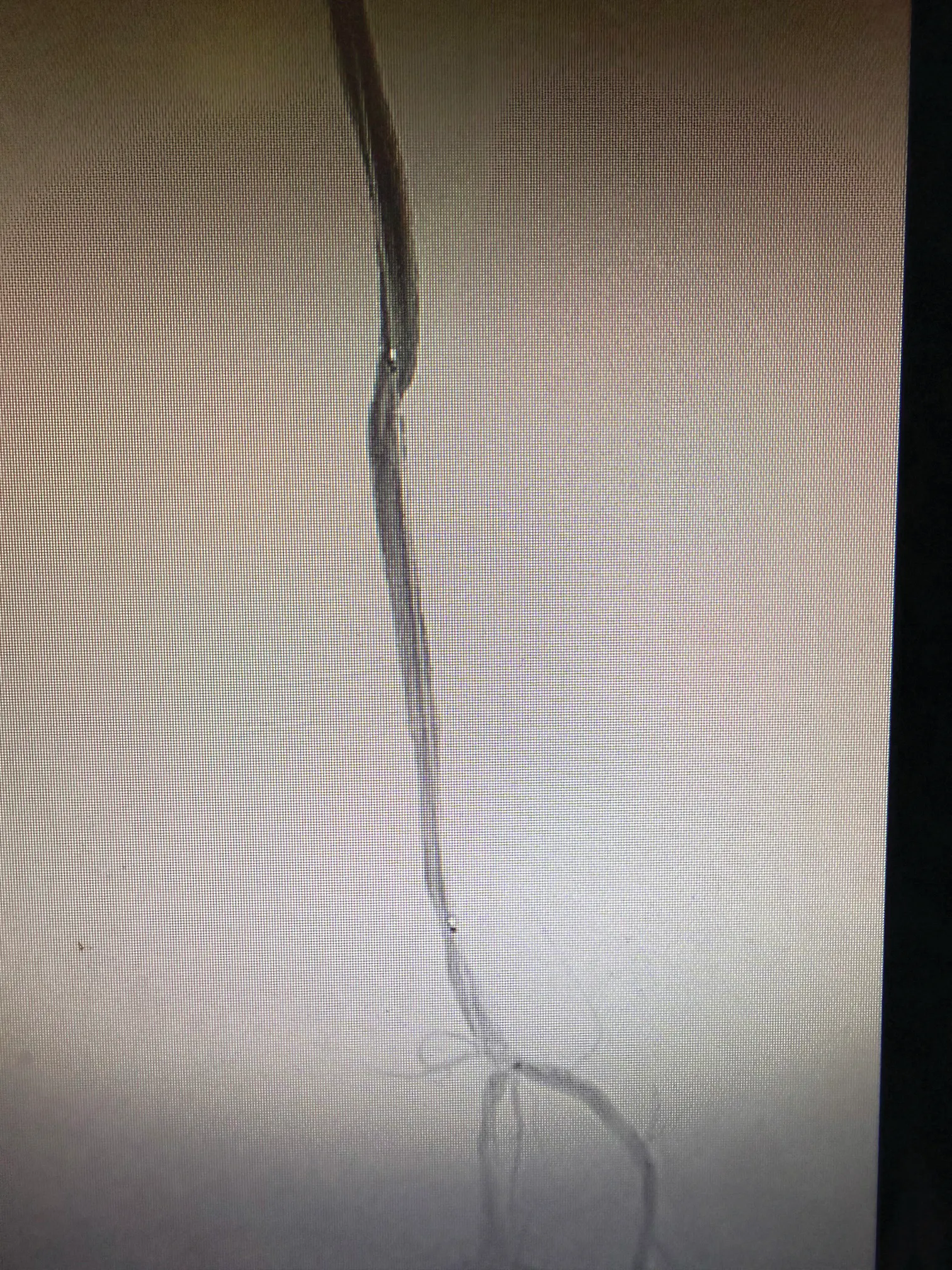
Figure 5. TORUS Stent Graft in the left popliteal artery with preserved runoff to the extremity.
POSTPROCEDURAL RESULTS
The patient was discharged 24 hours after the procedure, with an ABI of 1.0 (improved from 0.6 preprocedure). The patient has been followed for last 2 years and is doing well, with no symptoms of claudication pain in the left leg and a recent ABI of 0.97 in the left lower extremity.
*The DETOUR™ System is indicated for use for percutaneous revascularization in patients with symptomatic femoropopliteal lesions from 200 mm to 460 mm in length with chronic total occlusions (100 mm to 425 mm) or diffuse stenosis > 70% who may be considered suboptimal candidates for surgical or alternative endovascular treatments. The DETOUR™ System, or any of its components, is not for use in the coronary and cerebral vasculature.
The DETOUR™ System and associated components, ENDOCROSS™ Device and TORUS™ Stent Graft System, are not available in all countries or regions. Please contact your Endologix representative for details regarding product availability. Prior to use, refer to Instructions for Use for more information concerning Indications, Contraindications, Specific Anatomic Considerations, Warnings, Precautions, and Adverse Events. Rx only.
©2026 Endologix LLC. All rights reserved. MM3083-US Rev 01
Advertisement
Advertisement