Advertisement
Advertisement
September 2026
The Evolving Drug Elution Landscape in PAD
A global panel of experts explores the key developments and debates shaping the delivery of modern PAD care above and below the knee.
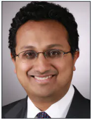
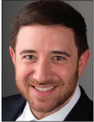
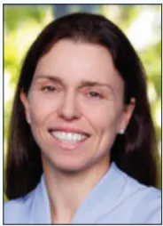
With Osamu Iida, MD; Joakim Nordanstig, MD, PhD; Sahil A. Parikh, MD;
Eric A. Secemsky, MD, MSc, RPVI, FACC, FAHA, FSCAI, FSVM; and Sabine Steiner, MD


BARE VERSUS PACLITAXEL VERSUS LIMUS
In your practice, how do you identify or predict the long-term potential for restenosis and repeat revascularization needs on a case-by-case basis, and how does that inform your decision-making?
Dr. Iida: In my practice, identifying the risk of restenosis relies on established systemic, limb, and anatomic factors. Systemically, I look at female sex, diabetes, chronic kidney disease, and a history of previous endovascular therapy. On a limb level, chronic limb-threatening ischemia (CLTI) naturally carries a higher risk than intermittent claudication (IC). Anatomically, factors like popliteal involvement, small vessel diameter, severe calcification, chronic total occlusion (CTO), and long lesions are key predictors. As an operator, when I encounter patients with a history of endovascular therapy, small vessels, severe calcification, or CTO, I proactively opt for drug-eluting devices because they offer significantly better long-term outcomes in these scenarios.
Prof. Nordanstig: I think the most accurate answer is that we are not yet able to reliably predict restenosis on the basis of clinical and morphologic characteristics alone. Several factors, including diabetes mellitus, chronic kidney disease, continued tobacco use, a history of restenosis, and systemic inflammation, have consistently been associated with an increased risk of restenosis. In addition, smaller vessel diameter, which is more common among women, increases the likelihood that restenosis will become clinically significant.
Several risk prediction models for restenosis have been proposed; however, to my knowledge, these remain largely investigational, although preliminary data are encouraging. Another promising area is molecular imaging. Vascular positron emission tomography, including fluorodeoxyglucose F18 and sodium fluoride F18 imaging, has the potential to identify metabolically active atherosclerotic plaque that may be at increased risk for restenosis. In the future, such techniques may facilitate more accurate risk stratification before lower extremity revascularization and support more individualized patient selection, particularly among patients with IC.
In my clinical practice, I perform close surveillance of all patients who undergo revascularization for CLTI, as restenosis in this population may have particularly serious consequences. I am liberal with using duplex ultrasound for postprocedural surveillance in CLTI. I monitor patients who have undergone infrapopliteal revascularization especially closely because restenosis rates are highest in this vascular territory; it’s not easy to detect up front; and evidence-based treatment options in this vessel segment remain comparatively limited, both at the time of the index procedure and after failure/occlusion of the initial revascularization.
Dr. Parikh: Restenosis is a function of lesion complexity including length, calcification, the presence of an occlusion, and patient risk factors (eg, diabetes and/or renal disease, among others). The more risk factors for restenosis, the greater the risk that a patient will have loss of patency and, therefore, the higher the value of providing antirestenotic therapy. We’ve learned this lesson in coronary artery disease over the past 30 years, and the same lessons apply to peripheral artery disease (PAD). As such, in my practice, I ask myself, “Why wouldn’t I use a drug-eluting technology?” in virtually every case. As a consequence, it is very rare that I would not use a drug-eluting therapy. The biggest reason I don’t is if we don’t have one available for that particular segment, as is the case for a below-the-knee (BTK) drug-coated balloon (DCB) for long-segment or inframalleolar (IM) disease. The absence of such therapy for the aortoiliac segment is less relevant, as the size of those vessels makes clinically meaningful restenosis unlikely.
Dr. Steiner: I consider response to vessel injury to be the main driver of restenosis. Lesion length is certainly an important predictor, as longer lesions mean a greater treated surface area and therefore a higher overall risk of restenosis, but I also look carefully at the underlying plaque morphology, degree of calcification, vessel diameter, lesion complexity, and, importantly, the quality of the final result after treatment.
I therefore try to assess not only the baseline anatomy but also how the vessel responds to intervention. Significant residual stenosis, dissection, recoil, or suboptimal vessel preparation can all indicate a higher risk of subsequent restenosis. In long and complex lesions, achieving an optimal acute result becomes particularly important. However, I ultimately put a strong emphasis on the long-term clinical outcome rather than focusing exclusively on patency. The potential need for repeat revascularization is influenced by the expected durability of the treatment but also by the patient’s symptoms and overall cardiovascular risk, functional status, and feasibility of future interventions.
In which patients and lesions do you ordinarily opt for a bare versus a coated device? Are there patients in whom you will categorically not use a drug delivery option?
Dr. Secemsky: In my practice, it’s rather infrequent to do an intervention without finishing with definitive drug-coated therapy. The exception is primarily in the anatomic locations where we do not have an approved device, such as in the iliacs and BTK in the case of DCBs. In a typical femoropopliteal (FP) case, I almost always use a drug device in some fashion, whether a balloon or a stent.
Dr. Iida: I generally default to drug-eluting devices. However, there are three specific scenarios where I avoid them. First, if a patient has a very short life expectancy (ie, < 1 year), they may not live long enough to experience the long-term benefits of drug elution. Second, for purely thrombotic lesions, the efficacy of drug delivery remains unclear. Finally, in CLTI patients with severely compromised IM runoff, specifically IM2, I avoid DCBs due to clinical concerns that downstream particulate embolization could acutely worsen the wound.
Dr. Parikh: As previously noted, I rarely use bare devices unless drug-eluting devices are not available for a particular segment. I suppose it’s reasonable to consider not using drug-eluting devices if patients absolutely cannot take antiplatelet therapy, but that’s relatively infrequent.
Dr. Steiner: In my practice, drug-eluting technology is the first-line strategy for most FP intervention treatment after appropriate vessel preparation. This is supported by current European Society of Cardiology (ESC) recommendations, the German Society for Vascular Medicine, and several expert consensus documents, reflecting the established benefit of drug delivery in reducing restenosis and repeat revascularization.1,2 I therefore use a DCB or drug-eluting stent (DES) in the vast majority of FP lesions. My choice is primarily determined by the anatomy and, importantly, the result after lesion preparation. I do not routinely select a bare device simply because of a particular patient characteristic. Non–drug-eluting devices are mainly an exception dictated by lesion-specific mechanical requirements. For example, in cases of extreme calcification requiring a pave-and-rack strategy, an interwoven stent may be necessary to achieve and maintain an adequate lumen.
Prof. Nordanstig: Recent randomized effectiveness trials of paclitaxel-coated devices (PCDs), including SWEDEPAD 1, SWEDEPAD 2, and BASIL-3, have not demonstrated the degree of clinical benefit that many had anticipated.3-5 As a trialist committed to evidence-based practice, these findings have made me more selective in my use of drug-coated devices. At the same time, I think the evidence remains clear that both paclitaxel- and limus-based devices reduce restenosis.
One possible explanation for the limited effect on limb salvage is distal embolization of paclitaxel crystals or excipients, a mechanism that may have been underestimated at least in CLTI. For this reason, I now use PCDs more cautiously in patients with CLTI. When restenosis prevention is a priority in the FP segment, I generally favor DESs over DCBs because they may carry a lower risk of distal embolization in my opinion.
The unresolved concerns regarding a possible long-term mortality signal with PCDs also make me reluctant to use these devices in younger patients with long life expectancy. Consequently, I do not routinely use PCDs for IC. This is consistent with current European Society for Vascular Surgery (ESVS) guidelines, which discourage their routine use during the index revascularization for IC while allowing consideration in selected cases of FP restenosis.6
Ultimately, device selection should be individualized and based on shared decision-making, with transparent discussion of both the established benefits in reducing restenosis and the remaining uncertainties regarding safety. In this context, limus-based technologies are a welcome development. They may offer a wider therapeutic safety margin, and advances in the balloon-coating technology itself have the potential to address some of the limitations of earlier-generation DCBs. Emerging effectiveness data, including findings from the SirPAD study,7 are encouraging, although further confirmation is warranted.
When do you opt for a DES versus a DCB once you’ve determined a drug-based option is ideal?
Dr. Steiner: I generally prefer a DCB when an adequate result can be achieved after vessel preparation and debulking, without significant residual stenosis, recoil, or flow-limiting dissection. Short, focal lesions are particularly well suited to this “leave-nothing-behind” approach, which preserves future treatment options. In contrast, longer lesions, significant recoil, post–percutaneous transluminal angioplasty dissections, suboptimal acute results, or heavily calcified segments often require additional mechanical support. In these situations, I have a lower threshold for using a newer-generation DES after adequate lesion modification.
The clinical presentation also matters. In patients with lifestyle-limiting claudication, I favor avoiding permanent implants whenever technically feasible, particularly when a durable DCB result can be achieved. In patients with CLTI, the threshold for definitive scaffolding is lower, as complex and heavily diseased lesions have a high likelihood of recoil or flow-limiting dissection, and a DES can provide a reliable and immediate mechanical solution.
Prof. Nordanstig: As noted, I now preferentially use DESs rather than DCBs for FP revascularization in patients with CLTI, given the potentially lower risk of distal paclitaxel crystal embolization. I believe this mechanism may be particularly detrimental in patients with advanced CLTI (Rutherford-Becker class 5 or 6). In the infrapopliteal circulation, I selectively use drug-eluting bioresorbable scaffolds (BRS), informed by the results of the LIFE-BTK trial,8 whereas I rarely use DCBs because randomized trials have not demonstrated consistent clinically meaningful benefit in this vascular territory. For FP in-stent restenosis (ISR), I selectively use DCBs when I consider the anticipated benefits to outweigh the potential risks. In Tosaka class 3 ISR lesions, I think that the evidence for using DCBs is less convincing than in less severe ISR scenarios.
Dr. Iida: Once I commit to a drug-based strategy, the choice between a DES and a DCB depends largely on the results of vessel preparation. In my experience, noncompliant balloons are essential for optimal prep. If we are left with residual stenosis or severe dissection, I will place a DES. Interestingly, I also compare the flow between the treated superficial femoral artery (SFA) and the deep femoral artery (DFA). If the DFA flow remains superior despite an angiographically acceptable SFA result, I will still consider a DES to maximize the hemodynamic gain.
Dr. Parikh: I’ll use stents when scaffolding is needed to optimize either dissections or luminal gain. The data for stents are strong, perhaps stronger than DCB in complex lesion subsets. However, when there is restenosis of stents, treatment is more difficult. The challenge is therefore to decide which patients will benefit most from scaffolds as opposed to balloon-based drug elution. As a practical standpoint, my threshold to use DES is lower in long segments and CTOs. With calcification, as long as I can get adequate expansion with a DCB or conventional DES, I will avoid use of a more purpose-built scaffold like Supera (Abbott), but there are many instances when I’ll use a DCB and Supera as a hybrid solution for calcific disease, especially at the adductor hiatus.
Dr. Secemsky: I employ a provisional stenting strategy. I typically perform the entire intervention from crossing to angioplasty and vessel prep in anticipation of using a DCB-first strategy, and provisional stenting as needed. My greatest DES usage is in long lesions and CTOs, as there are strong data supporting DES in these indications. But my overall goal is to limit the amount of permanent metal in the FP segment whenever possible.
If both are available to you, what will determine when you prefer a paclitaxel versus a limus device? Are there cases in which you’d avoid one or the other?
Dr. Parikh: I don’t think there’s compelling evidence in the above-knee circulation at present that limus is superior to paclitaxel. However, some early data demonstrating better durability of limus-based treatment is intriguing. In addition, the reduction in particulate embolization with limus DCBs in particular is also intriguing. In the end, I’d expect that superiority of long-term patency will drive our decision-making. In the absence of that, I think it’s a dealer’s choice. In the BTK circulation, the current data for limus devices, especially drug-eluting BRS, have made those devices seem superior, but it remains unclear if it’s the drug or the scaffold that is conferring that superiority. We’ll learn a lot more in the next couple of years.
Prof. Nordanstig: PCDs have a substantially more mature evidence base than limus-based technologies in PAD, particularly with respect to surrogate outcomes such as binary restenosis, target lesion revascularization (TLR), and late lumen loss. Clinical experience is also considerably greater (reflecting their longer use in PAD), although limus-eluting coronary stents have been used selectively for infrapopliteal ostial/close to ostial lesions for many years. That said, limus-based DCBs have the potential to address several of the limitations currently associated with paclitaxel-coated balloons. However, robust comparative effectiveness data remain limited. Additional randomized trials comparing limus-based balloons with uncoated balloons and PCDs are needed to determine whether this newer PAD technology offers noninferior or higher effectiveness, better safety, and, ultimately, greater patient benefit. In younger patients with high remaining life expectancy, it is especially important to do absolutely all I can to prevent the development of restenosis. Thus, I think limus-based therapy in these patients makes much sense, given the potentially larger safety margin that has been demonstrated in the coronary space.
Dr. Iida: Currently, there is no definitive borderline between paclitaxel and limus. Paclitaxel remains my first-line choice simply because of the extensive clinical evidence behind it. I typically reserve limus devices for cases of restenosis after previous paclitaxel treatment. However, there is a critical anatomic exception: If my guidewire tracking in a CTO goes subintimal, I am highly cautious about using paclitaxel due to the known risk of aneurysmal degeneration. From a mechanistic perspective, paclitaxel is cytotoxic, which can lead to tissue necrosis in the vulnerable subintimal space, whereas limus derivatives are cytostatic, which may offer a better safety profile in this regard. Therefore, in those specific subintimal cases, I will pivot to a limus device or even a nondrug strategy.
Dr. Steiner: In the FP segment, I still favor paclitaxel-based devices in most cases, given their extensive randomized trial evidence, established efficacy, and long-term clinical track record. At the same time, sirolimus is becoming an increasingly attractive alternative as new clinical data emerge. I would not categorically avoid either drug class. In my view, particularly in heavily calcified or fibrotic lesions, adequate vessel preparation and effective drug delivery are at least as important as the choice of drug itself. I would therefore not select a limus-based device solely on the basis of theoretical pharmacologic advantages. However, in cases where positive vascular remodeling or ectatic changes are a concern, particularly after previous paclitaxel-based treatment, sirolimus-based technologies may offer an attractive alternative given their distinct antiproliferative properties and potentially different effects on vascular healing. Overall, paclitaxel remains my default choice for now, but as longer-term clinical data on limus-based technologies mature, I expect the relative role of the two drug classes to become clearer.
BTK Applications
BTK treatment remains one of the most challenging frontiers for drug elution. Where do you see the greatest opportunities and limitations for drug-coated technologies in BTK intervention in the coming years?
Dr. Iida: Historically, the application of drug elution BTK has been limited to CLTI, primarily because of the poor initial success and long-term patency of plain balloon angioplasty. A significant opportunity here is that BTK-specific devices—designed with the appropriate pharmacokinetics and physical dimensions, rather than just being repurposed coronary or SFA devices—may help bridge this gap. DES can secure that crucial initial success, while DCBs or BRS can maintain long-term patency. If we can achieve this, we might see the indications expand to IC, as suggested by the 2024 ESC guidelines.1
However, the limitations remain significant. Pathologically, BTK disease is fundamentally different from the SFA; we are dealing with medial calcification and thrombotic occlusions in vessels < 1 mm. Furthermore, intervening in the IM space with severe medial artery calcification or small artery disease is often challenging with current atherectomy tools. Consequently, the future of BTK intervention may require us to carefully select patients who have a viable IM runoff bed to support the flow.
Dr. Steiner: BTK disease remains particularly challenging because recoil and severe calcification often play a major role in endovascular treatment failure, meaning that drug delivery alone may not be sufficient. In my view, one of the most exciting developments is the emerging evidence for drug-coated BRS, which combine antiproliferative drug delivery with temporary mechanical support to counteract recoil, without leaving a permanent metallic implant.
I see particular potential in a combination strategy using drug-coated BRS and DCB therapy. The scaffold can provide focal mechanical support and local drug delivery in segments where recoil is a major concern, while the DCB can treat the remaining lesion, including more distal segments where scaffolding may not be feasible. This approach could combine the advantages of mechanical support and antiproliferative therapy while minimizing permanent implants.
The evidence is still emerging, but I believe this combination approach could become an important direction for future BTK treatment, provided ongoing studies demonstrate durable clinical and limb-related benefits.
Prof. Nordanstig: In my opinion, this field has already taken an important step forward with the introduction of drug-eluting BRS. However, clinical evidence has thus far been largely limited to relatively short, noncalcified lesions, which differ substantially from contemporary practice, where many patients are elderly and present with extensive medial calcification, circumferential calcification, and long infrapopliteal occlusions. Nevertheless, the biologic rationale is compelling, and these devices should be evaluated in larger randomized trials enrolling broader, more representative patient populations. Partially absorbable drug-eluting scaffolds that adapt better to natural vessel movement and pulsatility compared with conventional stents are also promising. Devices like the DynamX Bioadaptor (Elixir Medical) are currently used in the coronary circulation, and I do think that this or similar such devices might hold potential in infrapopliteal vessels.
Next-generation limus-coated balloons are another promising development for infrapopliteal applications, although current evidence remains limited, and I believe additional randomized data are needed before their role can be more firmly established. More broadly, I also think the field should look beyond cytostatic and cytotoxic drug delivery. An alternative or complementary strategy may be to promote rapid endothelial regeneration after balloon angioplasty, thereby accelerating vascular healing (which in itself will prevent restenosis) rather than simply inhibiting neointimal hyperplasia.
Dr. Parikh: The numerous drug-eluting resorbable scaffold/BRS and DCB clinical trials with limus agents are very exciting. In addition, the sirolimus-coated Spur device (Reflow Medical) is also an intriguing drug delivery platform. We should see a lot of new data evaluating drug delivery in the coming year.
Outcomes and Endpoints
Historic and current clinical trials in PAD have primarily focused on various limb- and lesion-related outcomes, such as patency and revascularization success measures. Patient-centered (and -reported) outcomes are also important, although subjective data can be difficult to ascertain. Focusing on quality of life (QOL) in particular, which outcome measures do you find are most valuable? What are some of the challenges in accurately collecting these data in a PAD population and assessing technical approaches based on them?
Prof. Nordanstig: I agree that QOL outcomes remain underappreciated in vascular intervention, despite being among the outcomes that matter most to patients. There is also a common misconception that patient-reported outcome measures (PROMs) are inherently subjective or unreliable. In fact, modern QOL instruments are developed and validated using rigorous psychometric methods, including item response theory, and they undergo extensive testing to establish their validity, reliability, responsiveness, and sensitivity to change. In PAD, disease-specific instruments are generally preferable for evaluating treatment effects because they focus on symptoms and functional limitations directly attributable to PAD. By contrast, generic QOL measures may be less responsive, given the substantial burden of comorbidity in this population. The domains most relevant to PAD patients include physical function, pain, and social functioning, although their relative importance may differ between patients with IC and CLTI.
QOL analyses do present some methodologic challenges. Procedural placebo effects may overestimate treatment benefit in uncontrolled studies, and response shift—the adaptation of patients’ internal standards and expectations after a major life event such as serious illness—may lead to improved reported QOL time despite little change in disease status. However, both these principal limitations are adequately addressed by randomized trial design.
Among contemporary PAD-specific instruments, the 25-item and 6-item Vascular Quality of Life Questionnaire (VascuQoL) questionnaires are well validated. The abbreviated 6-item version is particularly attractive for routine clinical practice because it provides a concise yet robust assessment of disease-specific QOL. Especially in CLTI, it is my personal experience that more comprehensive, extensive questionnaires that include a wider range of items and questions are less well received and responded to, given the overall frailty of this elderly population. Data loss due to questions being left unanswered in an unrealistically long, complex questionnaire delivered to fragile patients is not desirable in scientific studies involving QOL data.
Dr. Parikh: PROMs and QOL data are very important for all technologies, especially in treating patients with IC where hard endpoint data are difficult to find. I think we need to look at the literature acquired in related fields, such as orthopedics, where joint replacement procedures are dictated by patient preference. We would stand to learn a lot by collaborating widely. Another challenge we have is the validation of these survey instruments in diverse patient populations. For example, a large segment of our population is Spanish speaking, and we do run the risk of missing data that are literally “lost in translation.” Culturally appropriate survey instruments that allow the synthesis of results across cultures need to be developed and more widely adapted for trial data to be adequate to facilitate decision-making.
Dr. Steiner: For me, walking capacity, functional status, symptom relief, and QOL are among the most valuable outcomes in PAD. In IC, the ability to walk further, become more active, and return to normal daily activities is ultimately more meaningful to the patient than patency alone. In CLTI, wound healing, pain, limb preservation, and maintaining independence are particularly important. At the same time, we should not underestimate the importance of patency and freedom from restenosis, particularly in CLTI. Restenosis can delay wound healing and cause recurrent ischemia, potentially putting the patient at increased risk of limb loss, even if this does not immediately translate into differences in amputation-free survival.
A major challenge is that PROMS are influenced by many factors beyond the treated lesion, including age, comorbidities, mobility, cardiovascular health, and psychosocial circumstances. This is particularly relevant in the highly heterogeneous CLTI population and makes it difficult to directly attribute changes in QOL to a specific technical intervention. Beyond revascularization, we also need to recognize that we are only beginning to understand how disease-modifying medical therapies may influence PAD progression and PROMs. Evidence that pharmacologic strategies can modify disease progression, and ultimately translate into improvements in PROMs, is still emerging. This further highlights the multifactorial nature of PROMs and the need to interpret them in the context of the overall patient journey, rather than attributing changes solely to the revascularization. Ultimately, I believe we need to combine objective measures of durability and limb outcomes with validated PROMs and functional assessments, while also understanding which factors truly matter most to patients.
Dr. Secemsky: Defining optimal endpoints is indeed challenging, especially for claudicants, who generally have a more benign overall course. In these patients, we are aiming to improve their QOL, but how we successfully measure this can be elusive. In clinical trials, patency has conventionally been considered an objective metric—is the target lesion open or not? But this may not necessarily be meaningful to the patient. As such, we have complemented this outcome with clinically driven target lesion revascularization, with the goal of tracking interventions driven by symptoms, but this endpoint is often critiqued, as it can be impacted by subjectivity.
QOL measures are also important ways to assess outcomes, and I am a big proponent of their inclusion in clinical trials. However, we need to acknowledge the limitations of PROMs. One major challenge is seen with QOL assessment across heterogeneous PAD populations. In trials and in the real world, patients' QOL can vary considerably, and their goals may look very different from one another. For instance, one patient may be comfortable spending their days on their couch, whereas for another, it is pain-free work days or running 5 miles.
We have seen QOL collected in PAD trials where discrepant findings are encountered. A good example of this is BEST-CLI, which was favorable for surgical revascularization primarily due to the need for less reintervention compared with the endovascular therapy arm. However, if the primary endpoint were QOL, the trial would have been marginally in favor of endovascular revascularization, which had modest improvements over surgery in regard to some QOL metrics at 1 year.
In a claudicant population, the severity of baseline symptoms is also critical, and changes in QOL are influenced by ceiling effects. For instance, in the IC arm of SWEDEPAD 2, 40% of randomized patients had Rutherford 1 or 2 symptoms. As such, their overall baseline QOL is only modestly impaired, leaving only so much room for improvement. For those who had Rutherford 3 symptoms, any QOL benefit will be more pronounced, as these patients have a larger potential range for improvement. QOL endpoints are also incredibly sensitive to how these data are collected.
In our center, we have a trained researcher sit in person with the patient and collect the patient's answers to these surveys. It is hard to do this by phone and can be affected by where the patient may be, how interested they are in participating, and other factors in their life that could be influencing their answers at that moment. Overall, it remains critical to include PROMs in trials, but due to this subjectivity and potential for bias, they are rarely a primary endpoint, especially when measured 1 year after a procedure.
Dr. Iida: QOL is a critical endpoint because the primary goal of revascularization in PAD is not necessarily to extend life but to restore the quality of it. If we use QOL as a primary endpoint, we must be extremely precise about who we are evaluating and what we are actually measuring. We have to strictly separate claudicants from CLTI patients. For a CLTI patient, a good QOL means maintaining a stable, wound-free status, avoiding major amputation, and staying out of the cath lab for reinterventions. It requires measuring mental well-being because the threat of amputation is a constant psychological burden. For claudicants, it’s much simpler: absolute walking distance.
When recent randomized controlled trials (RCTs), even those utilizing disease-specific tools like VascuQoL, suggest that drug-eluting devices do not improve QOL compared to bare devices, it often contradicts our real-world clinical experience. We see patients every day whose clinical courses are significantly improved because a drug-eluting device prevented restenosis.
I believe these trial results often stem from two primary methodologic issues. First is the insensitivity of current QOL tools to the benefit of avoiding reintervention. The primary advantage of drug elution is reducing TLR. Current questionnaires are often inadequate at capturing the psychological and physical relief a patient experiences by not having to undergo a repeat procedure. For a patient, staying out of the hospital is a major clinical benefit, but this episodic relief gets lost in standardized scoring. Second is the dilution effect caused by heterogeneous patient selection. If a trial pools patients with claudication together with CLTI patients or enrolls patients whose baseline QOL isn’t severely impaired to begin with, the true benefit of the device gets washed out. To accurately assess the value of drug-eluting technologies, we need endpoints that specifically weigh “freedom from reintervention” and capture the true mental burden of the disease.
The ESVS Guideline Update
How has your practice responded to or incorporated the findings of the SWEDEPAD 1 and 2 trials3,4 into its algorithms? And to the 2026 ESVS PAD guideline6 update?
Prof. Nordanstig: These trials have made me more selective in my use of PCDs. The benefits previously reported for reducing reinterventions were less pronounced in SWEDEPAD 1 and SWEDEPAD 2, the two largest randomized effectiveness trials in this field.3,4 In SWEDEPAD 1, no benefit was observed for harder true endpoints such as limb salvage or QOL. PCDs reduced reinterventions during the first year in CLTI, but this advantage was not sustained, suggesting that they delay rather than prevent repeat procedures. This might, of course, have patient value; but overall, it seems that previously reported benefits of these devices, mainly in claudication patients, did not translate to true benefits after CLTI revascularization.
SWEDEPAD 2 showed no significant treatment effect on any major endpoint studied, including PAD-specific QOL and hemodynamic metrics. The 1-year point estimate regarding target vessel reintervention rates did modestly favor PCDs. The latter neutral finding in SWEDEPAD 2 may, hypothetically, partly reflect Sweden's conservative approach to revascularization for IC, where symptomatic restenosis is often managed conservatively, with continued exercise therapy rather than repeated intervention. From a scientific standpoint, this endpoint might be less useful in patients with IC, at least in similar publicly funded health care systems like Sweden. Considering the SWEDEPAD 2 findings and the absence of convincing durable clinical benefit in long-term follow-up studies derived from a range of smaller randomized studies published after the previous ESVS guideline literature search, the guideline writing committee could not reasonably have recommended otherwise. Also considered was the still-unresolved late mortality signal that was replicated in SWEDEPAD 2. It should be emphasized that the ESVS guideline update only targeted claudication patients. Treatment safety is especially crucial as any revascularization indication remains relative in this patient population, and because there are other treatment options available (eg, vasoactive drugs, exercise).
Dr. Iida: In our practice, we have not fully adopted the SWEDEPAD findings or the 2026 ESVS guideline update into our algorithm. Paclitaxel remains the cornerstone of our practice. We have discussed this extensively, even with regulatory bodies like the Pharmaceuticals and Medical Devices Agency in Japan, and there is a shared understanding that downgrading a therapy to a class III recommendation based on a single RCT may be premature.
Dr. Steiner: I have not changed my treatment algorithm based on SWEDEPAD 1 and 2. Both trials have contributed valuable data to the ongoing discussion, but I do not consider them sufficient to fundamentally alter the role of drug-eluting technologies in my practice.
SWEDEPAD 2 was a large and important trial, but it was conducted in a single health care system and had several pragmatic limitations that may affect generalizability. These include the high proportion of patients with mild claudication (Rutherford 1-2, 40.4%); lack of standardization regarding devices, lesion preparation, and follow-up imaging; and registry-style follow-up without core lab assessment of patency or TLR. Importantly, the trial included a broad range of PCDs, which should not necessarily be considered equivalent in terms of efficacy and inhibition of restenosis. Given these limitations, I would not consider SWEDEPAD 2 sufficient on its own to fundamentally change my practice as proposed in the 2026 ESVS PAD guideline update. I therefore continue to use drug-eluting technologies.
Dr. Parikh: I disagree with the guideline update. I think it reflects poorly on the guideline process in general and ignores a lot of carefully conducted research with greater mechanistic underpinning.
Dr. Secemsky: SWEDEPAD follows a series of successful Swedish trials that have the ability to evaluate patients in registries and via health insurance databases. It is a unique infrastructure that provides valuable prospective data. As is the case with similar registries in the United States, we have to acknowledge the inherent limitations to these types of registry-based studies, particularly when attempting to perform comparative analyses that require detailed follow-up and adjudication of endpoints. This is particularly important for PAD trials. When assessing the success of a procedure or device, we often want to examine target lesion-related outcomes, which then allows us to associate these findings with clinical endpoints like symptom improvement. It becomes challenging to make this connection when a device trial does not include these anatomic-level data that should correlate with change in QOL.
In the United States, I have not seen a major shift in practice since SWEDEPAD and the ESVS guideline update. The mortality question has been carefully studied over more than 5 years by investigators, regulators, sponsors, and clinicians without concluding a direct relationship with decreased survival. These devices have been used in the United States for more than a decade now, and operators have had time to observe their patient outcomes. Like other therapies, there are many proponents who use these devices routinely, as well as nonadopters who never incorporated these devices into their practice, and there are people somewhere in between. Nonetheless, practice in the United States seems to be unchanged overall.
The guideline update was concerning to many of us in the United States, particularly those who have been involved in guideline development. Guidelines are not typically updated out of cycle, and do not use data from a single trial to change practice recommendations. While on the one hand we do not ever want to dismiss a safety signal, I am not aware of a previous trial using a QOL endpoint and secondary mortality data to change practice guidelines for well-proven therapies.
Among the more often cited criticisms of the 2018 Journal of the American Heart Association meta-analysis conclusions were the lack of identification of a causal link combined with a wide variety in causes of death.9 This was especially noteworthy given the established safety profile of paclitaxel administered in larger doses in oncology applications. The current findings from SWEDEPAD 2 also do not identify a causal link. How do you interpret the continued lack of a “smoking gun” despite extensive study of a potential late mortality signal? What is your understanding as to why there was a mortality signal at 5 years in one trial but not the other?
Dr. Steiner: I think the mortality signal deserves attention, but we still lack evidence for a causal relationship. There is no consistent dose-response relationship or convincing biological mechanism, and large population-based studies have not demonstrated a reproducible mortality signal. SWEDEPAD 2 itself was not primarily designed or powered to assess mortality, and with ongoing follow-up, the 5-year finding should be interpreted cautiously. The difference between SWEDEPAD 1 and 2 with respect to mortality is also difficult to explain and may reflect differences in patient populations and event rates, or simply statistical variation. Importantly, a signal emerging at one time point in one trial, without a consistent pattern across studies, does not establish causality. For me, this means we should continue long-term surveillance but avoid drawing conclusions that go beyond the available evidence.
Dr. Secemsky: It is challenging to interpret for a variety of reasons. It is hard to dismiss that we have seen a signal of harm between 2 and 5 years in both the original JAHA meta-analysis and in the claudication arm of SWEDPAD. However, we have never found a clear link to cause of death, nor any signals of specific differences in causes of death between those treated and not treated with drug-coated devices. Furthermore, we have numerous PCDs on the market now, ranging from arteriovenous fistula treatment to coronary intervention, and signals of harm have not been seen in data assessing these devices. Without a clear causal link, this remains a poorly explained signal that may be spurious and has not influenced survival rates in large population-based studies.
Prof. Nordanstig: Identifying a clear mechanistic explanation for the observed excess in late mortality associated with PCDs would be highly valuable, and it does remain frustrating that we have not yet been able to do so. However, I also believe we should remain open to the possibility that the association reflects a more generalized biological effect rather than a single, direct toxic mechanism. For example, persistent low-grade inflammation or immune modulation could plausibly contribute to a range of causes of death and represent the missing mechanistic link between local paclitaxel delivery and late mortality.
It is also important to recognize that the pharmacokinetics of the crystalline paclitaxel used on balloon- and stent-based platforms differ substantially from those of solvent-based paclitaxel formulations used in oncology. Consequently, safety data from systemic oncologic use cannot be directly extrapolated to endovascular drug-coated devices used in PAD.
Dr. Iida: The meta-analysis by Katsanos et al prompted extensive discussions across Japanese vascular and interventional societies. However, Japan has since generated robust, nationwide real-world data through our own registries consistently demonstrating the long-term safety of paclitaxel. Because we have our own solid evidence base, the reaction to the SWEDEPAD update has been more measured; we tend to rely on our accumulated data. Regarding the mortality signal, there are too many inconsistencies. Extracting a 5-year data point from a study with a much longer average follow-up, especially when the ultimate long-term data show no difference, is methodologically questionable. We use paclitaxel daily, and without a plausible biological mechanism—a “smoking gun”—it is difficult to justify restricting a highly effective therapy based on what is potentially an incidental finding.
The primary causes of death in patients with PAD and CLTI are cardiovascular events and infections. Do paclitaxel devices actually accelerate the progression of coronary or carotid artery disease? Does the use of paclitaxel compromise the immune system? While we certainly cannot completely ignore the mortality signal, from the perspective of the actual causes of death, the underlying mechanism remains entirely unclear. PCDs do have their drawbacks, but I believe they are safe tools provided we select DCBs with minimal downstream embolization and avoid deploying DCBs or DESs in the subintimal space.
Dr. Parikh: As the first author of the Lancet patient-level meta-analysis, we did not find a signal nor a plausible mechanism.10 I think this is, at best, a stochastic finding. I don’t think there is a plausible mechanism, a dose response, or a clear signal that should impact clinician behavior.
The accompanying editorial to the ESVS guideline states that there is no clear class effect across the heterogeneous current offering of PCDs and notes considerable heterogeneity in their associated data.11 However, the new recommendations regarding PCD usage do not delineate between different platforms, nor the overall classes of balloons and stents, when recommending when and in whom PCDs should be used. Can you address why this is?
Prof. Nordanstig: I was not involved in writing this editorial, but I personally would be cautious about concluding that there is no class effect at this stage. Direct comparisons among different PCDs are scarce, and the available head-to-head trials have generally shown similar efficacy, while lacking sufficient follow-up to meaningfully assess long-term mortality. Consequently, the guideline committee could not reasonably distinguish between individual devices, particularly in patients with IC, where the threshold for safety must be especially high.
SWEDEPAD, BASIL-3, and the many meta-analyses published so far have evaluated a range of PCDs. It remains possible that any potential mortality signal is device-specific rather than a class effect; however, SWEDEPAD 2 found no evidence to implicate any individual device.
Regarding the shared decision-making provision in the updated guideline, how should a care provider frame the disparate findings between the extensive FDA exploration, Centers for Medicare & Medicaid Services, and other major insurance/international database findings, and randomized trial reviews that did not find evidence of a mortality concern versus those of SWEDEPAD 2? And, how should SWEDEPAD 2’s own disparities in mortality signal based on the points of follow-up (ie, the finding appearing at 5 years and not at 7 years) be explained?
Prof. Nordanstig: This is a very important question. First, it should be emphasized that the observed excess in late mortality associated with PCDs has been modest, in SWEDEPAD 2 and in previous meta-analyses of industry-sponsored small pivotal RCTs. In SWEDEPAD 2, 104 of 565 patients in the paclitaxel group died within 5 years compared with 77 of 571 in the uncoated device group, corresponding to event rates of 4.57 and 3.28 deaths per 100 person-years, respectively.4 Differences of this magnitude are susceptible to residual confounding in observational studies, underscoring why RCTs remain the most reliable source of evidence for comparative safety.
It is equally important to recognize that SWEDEPAD 2 was not designed or powered to detect differences in mortality. The trial was initiated before the publication of the meta-analysis by Katsanos et al,9 with QOL as the primary outcome, not survival. The apparent attenuation of the mortality difference by 7 years should also be interpreted cautiously. As follow-up lengthens, all-cause mortality inevitably accumulates in both groups, and all Kaplan-Meier curves depicting mortality eventually converge if follow-up is long enough. Nevertheless, in SWEDEPAD 2, the separation between the survival curves from approximately 2 to 5 years closely mirrored the temporal pattern reported in the original 2018 Katsanos et al meta-analysis. Given this consistency, we did not believe that the finding could be dismissed, despite the uncertainty regarding its underlying mechanism.
Loss to follow-up, crossover, and lesion length have been identified as major confounders in previous studies. How were these elements addressed in SWEDEPAD 1 and 2?
Prof. Nordanstig: Both SWEDEPAD 1 and 2 achieved complete follow-up for mortality, a major strength of the registry-based randomized design. Linkage to the Swedish Population Register ensured complete ascertainment of deaths among all study participants.
We also performed several sensitivity analyses to assess the potential influence of treatment crossover and found no meaningful effect on the magnitude or direction of the mortality signal; these findings were presented at the Charing Cross Symposium earlier this year.12
Lesion length is an important but challenging variable to interpret, particularly in observational studies. Although longer lesions are associated with greater paclitaxel exposure, they also reflect more advanced atherosclerotic disease and, therefore, a higher baseline risk of death. In both SWEDEPAD trials, lesion complexity (including lesion length as defined by TransAtlantic Inter-Society Consensus II classification) was well balanced between the randomized treatment groups, minimizing the potential for confounding. This is entirely expected in trials of this size but exemplifies the power of randomization, especially in larger cohorts.
Numerous studies show efficacy of drug delivery in improved primary patency and TLR reduction. We understand that primary patency was not assessed in SWEDEPAD 1 and 2; what is your interpretation of why the reduction in TLR was relatively modest?
Prof. Nordanstig: As noted, SWEDEPAD 1 suggested that PCDs delay rather than prevent restenosis, since the reduction in target vessel revascularization (TVR) observed at 1 year was not sustained with longer follow-up. Nevertheless, the absolute reduction at 1 year was still potentially clinically meaningful: TVR occurred in 179 of 1,180 patients treated with PCDs and 224 of 1,175 treated with uncoated devices (hazard ratio, approximately 0.80).3 In my view, this effect is consistent with expectations for an unselected, real-world population with a range of lesion complexities. By contrast, many earlier pivotal trials enrolled highly selected patients with relatively short, stenotic, less calcified FP lesions and predominantly included patients with less severe symptoms than CLTI. As mentioned, the neutral findings in terms of target vessel reinterventions as well as overall rates of ipsilateral interventions in SWEDEPAD 2 may also speculatively reflect Sweden’s relatively conservative approach to repeat revascularization for claudication, where symptomatic restenosis is often managed without further intervention. In such settings, TLR or TVR may hence be a less informative measure of treatment effectiveness than patient-centered true outcomes, such as symptoms, functional status, and QOL. It is a different story in CLTI, as CLTI symptom relapse is a clearer indication for reintervention due to restenosis.
Forward Looking
From different drugs and excipient/coating technologies to delivery platforms ranging from balloons to permanent and transient scaffolds as well as direct delivery methods, iterative advancement continues across a variety of categories in the drug delivery field. What characteristic(s) would truly define a “next-generation” drug delivery device to you?
Dr. Secemsky: The continued evolution of drug delivery will involve factors that impact outcomes, including an improved understanding of optimal device sizing, ability to combine plaque modification with drug delivery, and improvement in drug transfer efficiency, particularly through the most challenging lesion subsets (ie, calcific disease). These factors are as important as any drug-coated device itself, and as we continue to iterate, we will likely see combination devices that can address many of these issues to maximize effective drug delivery.
Dr. Iida: A next-generation device should address two specific clinical challenges we face today. First, regarding paclitaxel DCBs, the powder-based coating presents a risk of downstream embolization, which can be harmful to the distal runoff. We need advancements in coating technologies that offer higher tissue transfer rates without the particulate downstream effect. Second, the issue of aneurysmal degeneration when paclitaxel is used in the subintimal space must be addressed. This is inherently a paclitaxel issue, which opens the door for limus-based or advanced nondrug therapies.
Ultimately, the core of intervention is stenting. If a peripheral DES emerges that achieves the level of success seen with coronary DES, it will become the mainstream. However, preserving the complex collateral networks in the leg is vital for limb function, which makes the leave-nothing-behind philosophy of DCBs highly relevant. I don’t believe we will see a complete shift to limus overnight; we need rigorous, head-to-head RCTs comparing limus and paclitaxel to truly define the next era of PAD care.
Dr. Steiner: To me, a truly next-generation drug delivery device would go beyond simply delivering a more effective drug or improving drug retention. The real breakthrough would be the ability to predict the biological response to vascular injury and tailor therapy accordingly. By integrating intravascular imaging with molecular plaque characteristics, we could potentially identify which lesions are prone to excessive neointimal hyperplasia or restenosis and determine which antiproliferative strategy, drug, dose, and duration of exposure would be most effective for that individual patient. Ultimately, I envision a shift from a “one-device-fits-all” approach toward personalized drug delivery based on lesion biology and the patient’s response to injury.
Dr. Parikh: There are a number of next-generation technologies that have been developed for vascular applications across the circulation, from cerebrovascular to coronary to peripheral. Each of these seeks to increase drug delivery efficacy and durability of impact while minimizing vessel toxicity. The ideal next-generation combinations will make drug delivery more reliable within a therapeutic window for a longer duration to reduce variability of effect. These combinations should also be pro-healing and antithrombotic. The biggest regulatory challenge continues to be the ability to navigate the clinical-regulatory pathways for combined drug-device combinations, where new drugs coming into the drug-eluting device space have to navigate first the drug pathways and then the combined drug-device pathways prior to coming to market. At present, that pathway is fiscally untenable and has led to a restriction of innovation in this field. If we can figure that out, I’m sure our clinician-scientists can bring forward meaningful innovation over the next decade.
Prof. Nordanstig: One promising but poorly explored direction is to expand beyond conventional antiproliferative drug coatings. Agents that promote endothelial repair and vascular healing, rather than simply inhibiting neointimal hyperplasia, may provide a more favorable balance between restenosis prevention and vascular recovery. Both paclitaxel- and limus-based therapies delay endothelial healing, which may prolong the period of thrombotic risk and, in the case of stents, delay device integration. Limus-based therapies are indeed mechanistically favorable in this context due to their broader therapeutic safety margin.
I also believe that greater collaboration between clinicians, engineers, and industry is needed to improve coating technologies and other aspects of the device beyond the actual drug(s) delivered. More efficient local drug delivery with minimal downstream embolization may be particularly important in patients with CLTI, where distal perfusion and microvascular reserve are already severely compromised. This consideration is one reason why I currently favor DESs over DCBs in selected patients, although permanent metallic implants do have well-recognized limitations long term. Drug-eluting BRS offer an attractive alternative in this context by combining transient mechanical support with local drug delivery during the period when it’s most needed, while avoiding the long-term disadvantages of a permanent implant. Still, these have yet to show effectiveness in patients with more complex lesions and other vessel segments beyond the infrapopliteal, and they may be restricted by limited radial strength. Likewise, interwoven nitinol stents have improved the treatment of complex FP lesions, and the development of an interwoven drug-eluting platform represents a logical next step.
In the end, I cannot really postulate a single perfect device, and I do think we need a range of more effective and safer devices, so that we can tailor treatment to individual cases based on comprehensive evaluation of the detailed characteristics of the lesions we engage with.
1. Mazzolai L, Teixido-Tura G, Lanzi S, et al; ESC Scientific Document Group. 2024 ESC guidelines for the management of peripheral arterial and aortic diseases. Eur Heart J. 2024;45:538-3700. doi: 10.1093/eurheartj/ehae179
2. Malyar N, Behrendt CA, Espinola-Klein C, et al. S3-Leitlinie zur diagnostik, therapie und nachsorge der peripheren arteriellen verschlusskrankheit. Vasa. 2025;54:1-119. doi: 10.1024/0301-1526/a001171
3. Falkenberg M, James S, Andersson M, et al; SWEDEPAD trial investigators. Paclitaxel-coated versus uncoated devices for infrainguinal endovascular revascularisation in chronic limb-threatening ischaemia (SWEDEPAD 1): a multicentre, participant-masked, registry-based, randomised controlled trial. Lancet. 2025;406:1103-1114. doi: 10.1016/S0140-6736(25)01585-5
4. Nordanstig J, James S, Andersson M, et al; SWEDEPAD trial investigators. Paclitaxel-coated versus uncoated devices for infrainguinal endovascular revascularisation in patients with intermittent claudication (SWEDEPAD 2): a multicentre, participant-masked, registry-based, randomised controlled trial. Lancet. 2025;406:1115-1127. doi: 10.1016/S0140-6736(25)01584-3
5. Bradbury AW, Hall JA, Popplewell MA, et al; BASIL-3 investigators. Plain versus drug balloon and stenting in severe ischaemia of the leg (BASIL-3): open label, three arm, randomised, multicentre, phase 3 trial. BMJ. Published online February 24, 2025. doi: 10.1136/bmj-2024-080881
6. Nordanstig J, Hinchliffe R, Lejay A, Behrendt CA; ESVS Peripheral Arterial Disease Guidelines Writing Committee; ESVS Guidelines Steering Committee. Editor’s choice–focused update on paclitaxel coated technologies, from the 2024 European Society for Vascular Surgery (ESVS) guidelines on the management of asymptomatic peripheral arterial disease and intermittent claudication. Eur J Vasc Endovasc Surg. 2026;71:923-927. doi: 10.1016/j.ejvs.2026.04.037
7. Barco S, Engelberger RP, Held U, et al; SirPAD investigators. Sirolimus-coated balloon angioplasty for infrainguinal artery disease. N Engl J Med. 2026;395:561-570. doi: 10.1056/NEJMoa2600360
8. Varcoe RL, DeRubertis BG, Kolluri R, et al; LIFE-BTK investigators. Drug-eluting resorbable scaffold versus angioplasty for infrapopliteal artery disease. N Engl J Med. 2024;390:9-19. doi: 10.1056/NEJMoa2305637
9. Katsanos K, Spiliopoulos S, Kitrou P, et al. Risk of death following application of paclitaxel-coated balloons and stents in the femoropopliteal artery of the leg: a systematic review and meta-analysis of randomized controlled trials. J Am Heart Assoc. 2018;7:e011245. doi: 10.1161/JAHA.118.011245
10. Parikh SA, Schneider PA, Mullin CM, et al. Mortality in randomised controlled trials using paclitaxel-coated devices for femoropopliteal interventional procedures: an updated patient-level meta-analysis. Lancet. 2023;402:1848-1856. doi: 10.1016/S0140-6736(23)02189-X
11. Behrendt CA, Bertges DJ. What are the indications for drug eluting technology in peripheral artery disease management? Eur J Vasc Endovasc Surg. 2026;71:918-920. doi: 10.1016/j.ejvs.2026.02.047
12. Nordanstig J. The SWEDEPAD trial update: Subgroup analyses and completing risk assessment. Presented at: Charing Cross Symposium; April 21-23, 2026; London, United Kingdom.
Disclosures
Dr. Iida: Unavailable at the time of publication.
Prof. Nordanstig: Principal Investigator, SWEDEPAD; Chair, ESVS Guideline Committee.
Dr. Parikh: Advisory board for Abbott, Boston Scientific, Cordis, Medtronic, Philips, Rheoxtech, Stryker, W.L. Gore; research for Abbott, Acotec, Akura Medical, Boston Scientific, Concept Medical, FastWave Medical, J&J Medtech/Shockwave, Reflow Medical, and Regeneron; consultant to Evident Vascular, J&J MedTech/Shockwave, Terumo, Penumbra, and Eli Lilly; equity in Advanced NanoTherapies, EnCompass Vascular, and R3 Vascular.
Dr. Secemsky: Consultant to Abbott, AngioDynamics, Asahi, BD, Bayer, Boston Scientific, Conavi, Concept Medical, Cook Medical, Cordis, Endovascular Engineering, Evident Vascular, GE, Gore, Haemonetics, Medtronic, Nipro, Penumbra, Philips, RapidAI, Rampart IC, R3, Recor, Regeneron, Shockwave, Siemens, SoniVie, Teleflex, Terumo, Thrombolex, VentureMed, Verve, Zoll.
Dr. Steiner: Consultant to AngioDynamics, Abbott, Becton Dickinson, Bentley, Boston Scientific Corporation, Cook Medical, Cordis, Medtronic, Novartis, Reflow Medical, Shockwave, and Teleflex.
Advertisement
Advertisement