Advertisement
Advertisement
July 2025
Sponsored by Penumbra, Inc.
Transforming VTE Interventions: Lightning Flash™ 2.0—Speed, Safety, Simplicity in Action
With Massimo Pieraccini, MD; Pedro Pardo Moreno, MD, PhD; Juan José Ciampi Dopazo, MD; Gonzalo Ruíz Villaverde, MD; Pablo Navarro Vergara, MD; and Emma Wilton, MD
Case 1: Intermediate-High–Risk PE

Massimo Pieraccini, MD
Interventional Radiology Department
Ospedale Misericordia
Grosseto, Italy
CASE PRESENTATION
A female hypertensive patient in her mid-90s presented with progressive dyspnea and signs of acute respiratory failure and congestive heart failure. Arterial blood gas analysis showed hypoxemia and hypocapnia. High-sensitivity cardiac troponin T was elevated (65 ng/L), suggestive of myocardial strain. D-dimer was not performed due to the high clinical suspicion of pulmonary embolism (PE).
CT pulmonary angiography (CTPA) revealed bilateral thromboembolic occlusion originating from the main pulmonary arteries (PAs), extending to lobar and segmental branches of all lobes (Figure 1).
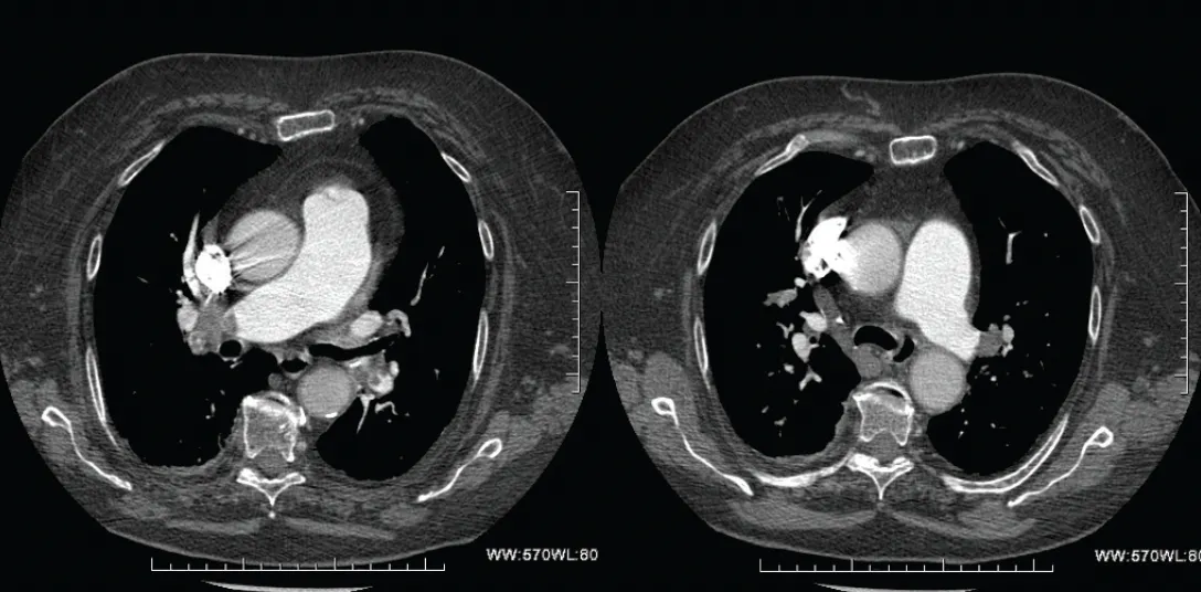
Figure 1. CTPA showing bilateral PE.
Transthoracic echocardiography showed preserved left ventricular (LV) function (ejection fraction, 55%) with concentric hypertrophy. The right atrium and ventricle were dilated (right ventricular [RV] diameter, 40 mm), with reduced RV systolic function (RV fractional area change, 25%; tricuspid annular plane systolic excursion [TAPSE], 14 mm) and a positive McConnell sign. Estimated systolic PA pressure (sPAP) was moderately elevated.
The patient was classified as intermediate-high–risk PE (PE Severity Index class III), with evidence of RV dysfunction and myocardial injury, although she was hemodynamically stable.
INTERVENTION
Given the high thrombotic burden, impaired RV function, and the patient’s frailty (limiting systemic thrombolysis), a mechanical thrombectomy approach was chosen.
A 16-F, 33-cm Gore® DrySeal sheath (Gore & Associates) was inserted via the right common femoral vein (CFV). PA catheterization with an angled pigtail catheter allowed initial pressure measurement (sPAP: right, 55 mm Hg; left, 43 mm Hg). A Lightning Flash™ 2.0 aspiration catheter (Penumbra, Inc.) was advanced easily into the right PA. Targeted thrombectomy was performed under fluoroscopic guidance, followed by selective angiography and repeat pressure measurements (Figure 2). The same procedure was conducted on the left PA (Figure 3). Total blood loss was approximately 80 mL, and procedure time was 35 minutes. Postprocedure hemodynamics showed significant improvement (sPAP: right, 33 mm Hg; left, 29 mm Hg).
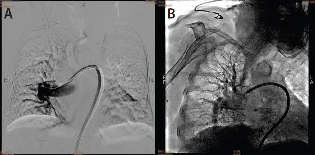
Figure 2. Pretreatment (A) and post-treatment (B) pulmonary angiography of the right PA.
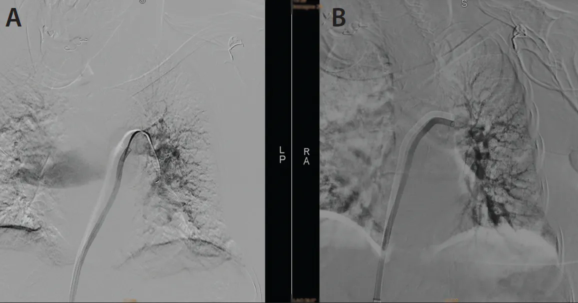
Figure 3. Pretreatment (A) and post-treatment (B) pulmonary angiography of the left PA.
POSTPROCEDURE FOLLOW-UP AND OUTCOMES
Repeat echocardiography showed normalization of RV function with TAPSE improved to 18 mm and RV diameter reduced to 38 mm. No periprocedural complications were noted. The patient experienced rapid clinical recovery with marked improvement in oxygenation and dyspnea.
DISCUSSION
This case highlights the safety and efficacy of percutaneous mechanical thrombectomy with Lightning Flash in patients with acute intermediate-high–risk PE and RV dysfunction. The navigability of the system was excellent, with smooth and intuitive advancement into both right and left PAs. The flexibility of the catheter shaft, combined with precise torque response, facilitated accurate positioning in the segmental branches.
In addition, Penumbra’s CAVT™ Computer Assisted Vacuum Thrombectomy allowed us to monitor aspiration effectiveness in real time, enabling rapid and efficient aspiration cycles and significantly reducing overall procedural time and blood loss, which was limited to approximately 80 mL.
Despite her age, the patient tolerated the procedure well and showed rapid hemodynamic and echocardiographic recovery. In fragile or anticoagulation-constrained populations, mechanical thrombectomy with Lightning Flash 2.0 can be an effective and a safe therapeutic strategy for acute PE, offering a valuable alternative to systemic thrombolysis or anticoagulation alone. A multidisciplinary evaluation and tailored risk stratification are essential for optimizing outcomes.
Case 2: High-Risk PE

Juan José Ciampi Dopazo, MD
Interventional Radiology Department
Hospital Universitario Virgen de las Nieves
Granada, Spain
Gonzalo Ruíz Villaverde, MD
Interventional Radiology Department
Hospital Universitario Virgen de las Nieves
Granada, Spain
Pablo Navarro Vergara, MD
Interventional Radiology Department
Hospital Universitario Virgen de las Nieves
Granada, Spain
CASE PRESENTATION
A male patient in his early 70s was brought to the emergency department with two syncopal episodes with vegetative cortex at home. He presented with sudden-onset dyspnea without chest pain. At the time of home care, the patient’s blood pressure (BP) was 90 mm Hg and heart rate (HR) 104 bpm. Electrocardiography showed ST-segment depression in II, III, aVF, and V3 to V6. SpO2 was 95% (FiO2, 24%).
Suspecting acute pulmonary thromboembolism, CTA and echocardiography were performed.
CTA showed acute bilateral pulmonary thromboembolism with significant thrombotic burden and radiologic signs of right heart overload (Figure 1). The main PA trunk was at the upper limit of normal (29 mm). The ascending aorta was within normal limits (32 mm). Rectification of the interventricular septum with inversion of the RV/LV ratio was noted.
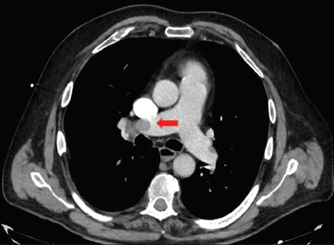
Figure 1. CTPA upon admission showing complete occlusion of the right PA (red arrow).
Echocardiography showed a severely dilated right ventricle with depressed systolic function, dilated right atrium, and moderate pulmonary hypertension. The left ventricle had normal dimensions and ejection fraction. TAPSE was 13 mm, and estimated PAP was 43 mm Hg. The inferior vena cava (IVC) was dilated with decreased inspiratory collapse. At this time, the patient was classified as having high-risk acute PE.
INTERVENTION
In cases of high-risk PE, the first treatment option is systemic thrombolysis, with thrombectomy recommended as a rescue treatment or as an alternative in cases of absolute contraindication for thrombosis.
In our case, fibrinolysis was ruled out due to clinical stabilization (BP, 131/57 mm Hg; HR, 97 bpm) in the hospital emergency department. Anticoagulant therapy was initiated, with no response after 12 hours of hospitalization. Given the characteristics of the case and aiming for an immediate re-patency, it was decided to perform PA thrombectomy with Lightning Flash 2.0.
The right PA was accessed, revealing complete occlusion of the right PA (Figure 2). The thrombectomy was then performed through a 16-F Performer introducer (Cook Medical) using a 16-F Lightning Flash 2.0 aspiration catheter, making two passes.
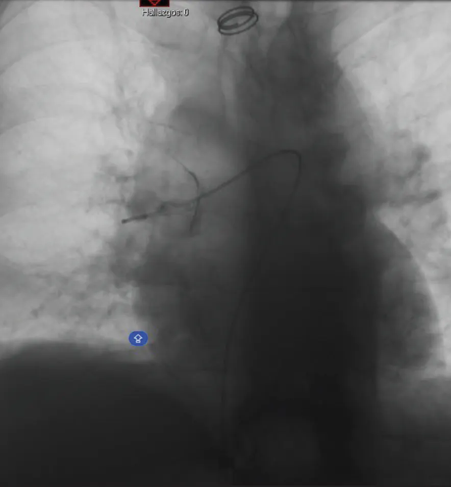
Figure 2. Selective arteriography of the right PA (complete occlusion).
Final pulmonary angiography revealed an approximately 90% decrease in thrombotic burden compared to initial angiography (Figure 3 and Figure 4). The procedure proceeded uneventfully.
After thrombectomy, SpO2 was 96%, BP was 180/90 mm Hg, and HR was 80 bpm.
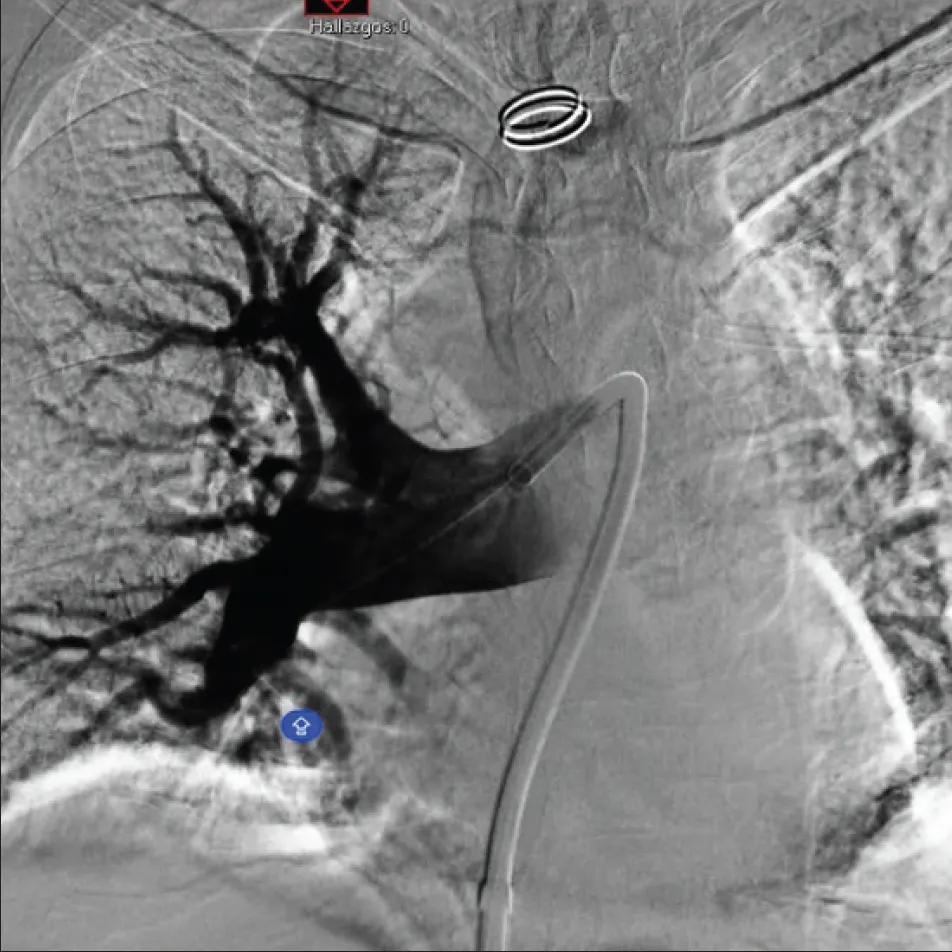
Figure 3. Post-treatment pulmonary angiography of the right PA.
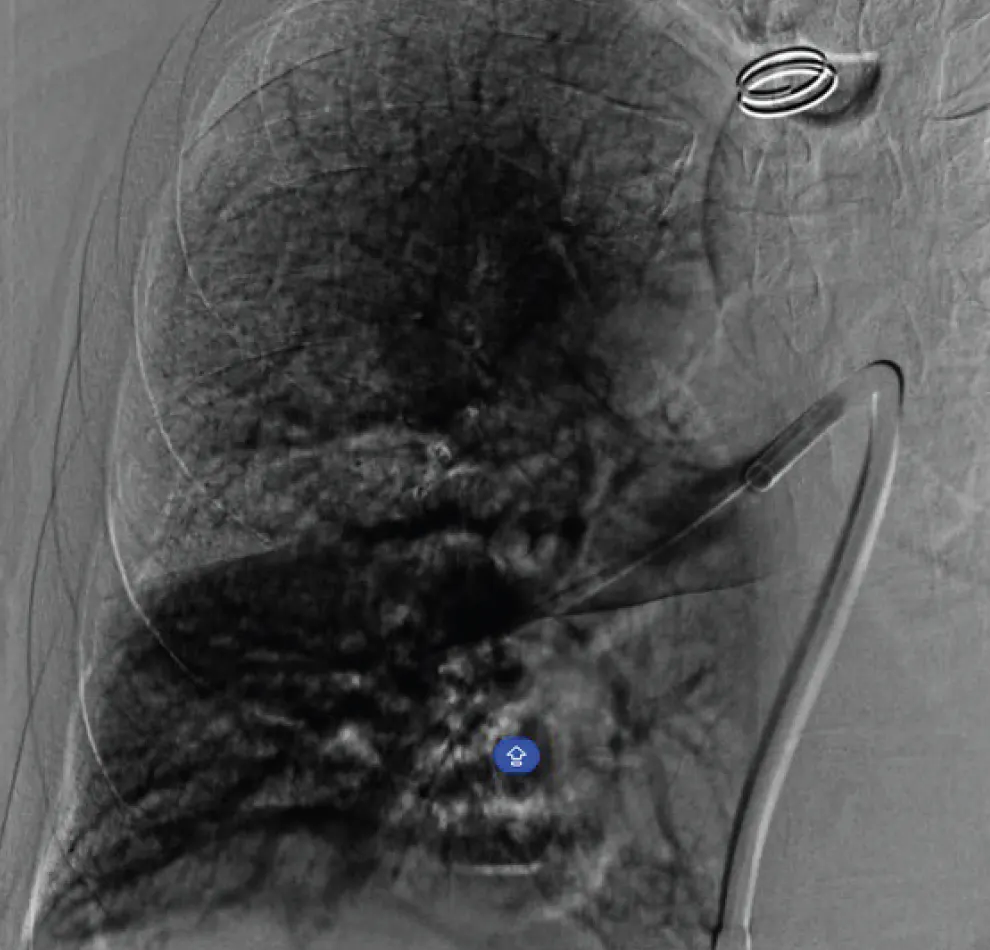
Figure 4. Final parenchymogram.
POSTPROCEDURE FOLLOW-UP AND OUTCOMES
In the 48 hours following the procedure, we routinely performed clinical follow-ups, CTA, and echocardiography on all patients. The post-thrombectomy CTA showed decreased post-treatment thrombotic burden with disappearance of the thrombus from the main branches and trunk, with partial thrombosis persisting in the lobar branch for the left upper lobe, right upper lobe, and subsegmental thrombosis in the right lower and left lower lobes (Figure 5).
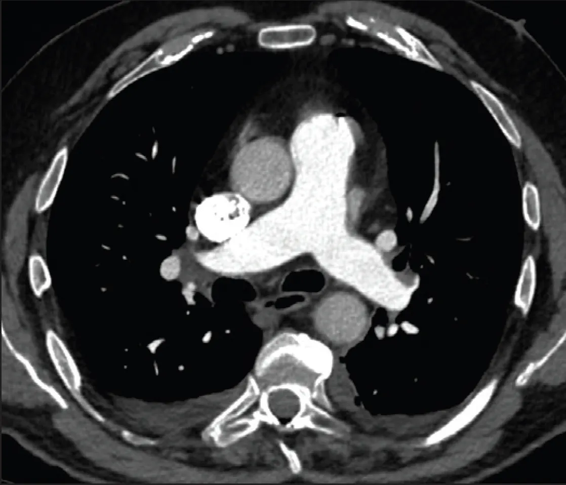
Figure 5. CTPA at 48-hour follow-up.
Post-thrombectomy echocardiography revealed a left ventricle with normal dimensions and wall thickness, without segmental contractility defects, and preserved ejection fraction (visual, 60%-65%). The right ventricle was slightly dilated with normal ejection fraction. TAPSE was 21 mm, and estimated PAP was normal. The IVC was normal, with mild tricuspid regurgitation. There was mild mitral regurgitation, and the left atrium was dilated.
After percutaneous thrombectomy and initiation of anticoagulation with low-molecular-weight heparin, the patient showed favorable outcomes, improving clinical symptoms and radiologic, laboratory, and echocardiographic findings observed on admission. At discharge, the patient was eupneic and tolerated light physical activity.
DISCUSSION
In our experience, pulmonary thrombectomy with the Lightning Flash appears to be a safe and efficient procedure for the treatment of PE in high- and intermediate-high–risk patients, with a low complication rate. Lightning Flash 2.0 provides CAVT, designed for accelerated thrombus detection and removal. Thromboaspiration capacity is similar to larger catheters while maintaining a lower profile designed for the pulmonary anatomy with excellent navigability.
Case 3: Deep Vein Thrombosis

CASE PRESENTATION
A female patient in her mid-50s was diagnosed with acute left iliofemoral vein thrombosis. She had a 13-day history of left lower abdominal/flank pain, venous claudication at short distances, and pain and swelling of the left lower limb. She had no significant past medical history. On examination, the left iliac fossa was tender with swelling and discoloration of the left lower limb. There was no phlegmasia. She had been started on anticoagulation. A CT scan had revealed lower IVC and left iliac vein acute thrombosis with inflammatory changes. She was planned for IVC/left iliac vein thrombectomy with the possibility of iliac venous stenting. Despite anticoagulation, her symptoms deteriorated. A preoperative venous duplex ultrasound showed thrombus propagation into the left CFV, femoral vein (FV), profunda vein (PFV), popliteal vein (PV), and upper tibial veins.
TREATMENT OPTIONS
At the time of intervention, it was 20 days following onset of symptoms. Mechanical thrombectomy was an option; however, with the propagation of thrombus into the tibial vessels, we decided to carry out aspiration thrombectomy with the Lightning Flash 2.0. This enabled us to clear the acute thrombus in the inflow veins, including the tibial veins.
INTERVENTION
Once in the interventional radiology suite, the patient was positioned supine. The procedure was performed under general anesthesia. Thromboembolic deterrent stockings and intermittent pneumatic compression boots were applied. These remained on until the patient began mobilizing. The right groin and left upper thigh were prepped and access was achieved under ultrasound guidance into the right CFV and left great saphenous vein (GSV). A 16-F Gore DrySeal sheath was inserted into left GSV. Initial venography confirmed occlusive thrombus within the lower IVC, left iliac vein, CFV, FV, PFV, PV, and upper tibial veins (Figure 1). Temporary embolic protection was placed in the infrarenal IVC from the right CFV access. Heparin boluses were given, with a target activated clotting time > 250 seconds. Thrombectomy of the IVC and left iliac vein was performed with the Lightning Flash 2.0 from left GSV access (Figure 2). A tight May-Thurner compression of the left common iliac vein (CIV) was noted. An up-and-over retrograde thrombectomy was then performed to remove thrombus in the left PFV, FV, and PV via right CFV access (Figure 3). Good thrombus clearance was achieved. The left CIV compression was treated with venoplasty to 14 mm, and a 14- X 150-mm Abre stent (Medtronic) was deployed from the ipsilateral GSV access. Manual compression only was required for access site hemostasis.
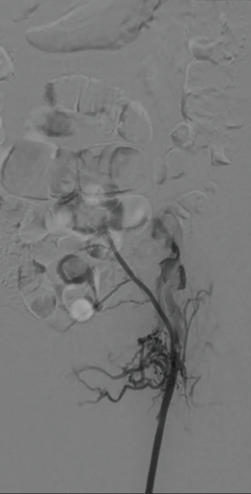
Figure 1. Initial venogram showing occlusive thrombus in the left iliac and CFV.
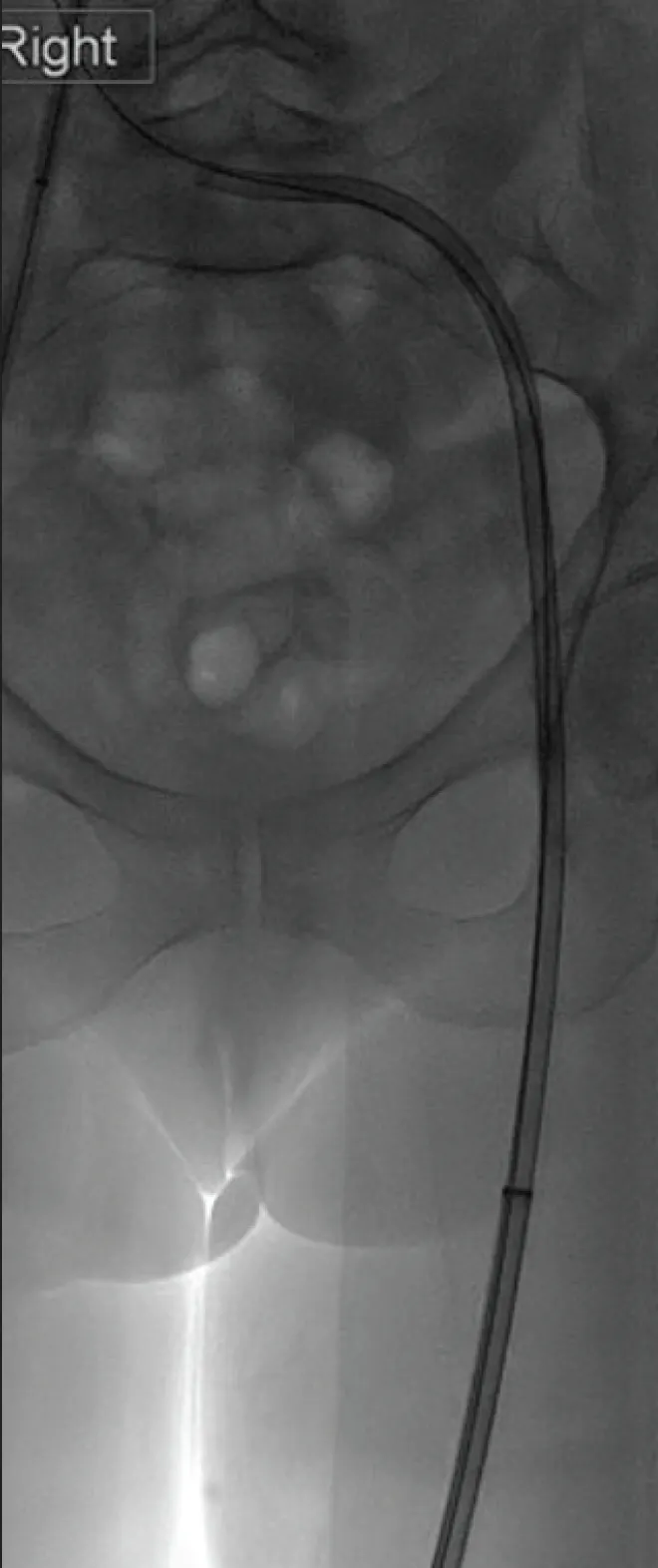
Figure 2. The Lightning Flash 2.0 aspiration catheter from the left GSV access.
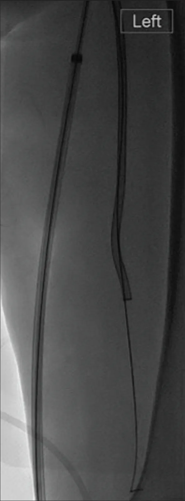
Figure 3. The Lightning Flash 2.0 aspiration catheter from the right CFV access (upsized to 16-F Gore DrySeal sheath) to clear the left CFV, FV, PFV, and PV.
POSTPROCEDURE OUTCOMES
Final imaging confirmed patency of the left iliac vein stent and treated inflow veins (Figure 4). The patient tolerated the procedure well. She was discharged home the following day on split-treatment-dose low-molecular-weight heparin, as per our protocol, and wearing compression stockings. Her symptoms had improved, and duplex ultrasound confirmed stent patency. Our standard stent surveillance was organized. Stent and inflow vessel patency has remained to date (> 6 months follow-up).
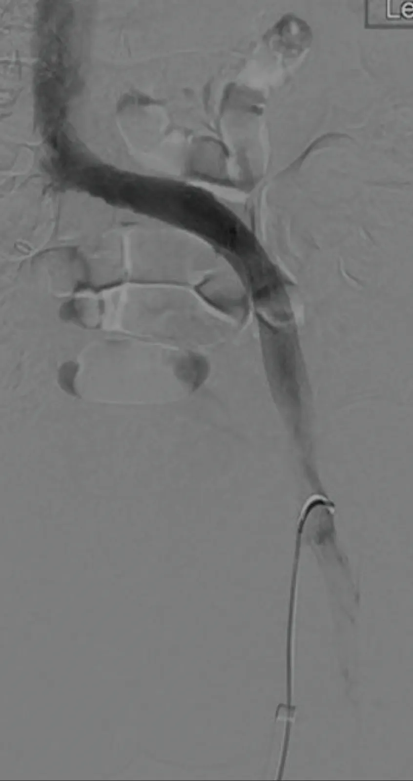
Figure 4. Final venogram showing the stented left May-Thurner compression point and good thrombus clearance.
DISCUSSION
Optimizing deep venous inflow during thrombectomy is essential to maintain vein and stent patency.1-3 This case employed a dual-access technique to aid inflow vein clearance.4 Mechanical thrombectomy alone could have been performed in this case; however, the thrombus extension into the tibial veins made CAVT preferable. According to our experience, the Lightning Flash 2.0 also allows treatment of patients with more subacute thrombus (> 14 days from onset of symptoms) due to the 16-F catheter. The procedure time for this case was only 90 minutes due to the ease of use of the device and shortening the aspiration time required. We have not had any significant complications using the Lightning Flash 2.0 via a 16-F sheath in a superficial vein of appropriate size and use either manual compression only or a purse-string suture for hemostasis.
Acknowledgment: Dr. Andrew Wigham, Consultant Interventional Radiologist, Oxford University Hospitals NHS Foundation Trust, was involved in the treatment decision-making and performance of the procedure.
Dr. Pieraccini, Dr. Pardo Moreno, and Miss Wilton were compensated in association with this article.
Disclaimer: The opinions and clinical experiences presented herein are for informational purposes only. The results may not be predictive of all patients. Individual results may vary depending on a variety of patient-specific attributes.
1. Grøtta O, Enden T, Sandbæk G, et al. Infrainguinal inflow assessment and endovenous stent placement in iliofemoral post-thrombotic obstructions. CVIR Endovasc. 2018;1:29. doi: 10.1186/s42155-018-0038-9
2. Yu Q, Kwak D, Tran P, et al. Single-session thrombectomy without thrombolysis for iliofemoral deep vein thrombosis: a single-center experience of ClotTriever with evaluation of predictors for reocclusion. The Arab J Intervent Radiol. 2024;8:19-26. doi: 10.1055/s-0043-1775992
3. Jalaie H, Barbati ME, Piao L, et al; International CVO Classification Study Group. Prognostic value of a classification system for iliofemoral stenting in patients with chronic venous obstruction. Eur J Vasc Endovasc Surg. 2025;69:315-322. doi: 10.1016/j.ejvs.2024.10.002
4. Wise R, Qamhawi Z, Wilton E, Wigham A. Iliofemoral DVT thrombectomy: a dual-access approach to optimise inflow. CVIR Endovasc. 2025;8:43. doi: 10.1186/s42155-025-00543-0
Advertisement
Advertisement