Advertisement
Advertisement
August 2026
Sponsored by Inquis Medical, Inc.
Simplifying PE Treatment: Real-World Experience With the AVENTUS® Thrombectomy System
With Austin Bourgeois, MD; Thomas Wong, MD; Ripal T. Gandhi, MD, FSVM, FSIR; and Patrick Muck, MD
Early Experience With the AVENTUS® Thrombectomy System for PE: Observations on Tissue Sensing, Directional Aspiration, and Integrated Blood Reinfusion
By Austin Bourgeois, MD
Mechanical thrombectomy (MT) has become a key therapeutic modality for patients with intermediate- and high-risk pulmonary embolism (PE). Aspiration-based platforms effectively reduce thrombus burden and improve hemodynamics, and they may reduce mortality in appropriately selected patients. As the field has evolved, medical device development has focused on improving clot extraction efficiency while minimizing blood loss, simplifying procedural workflows, and expanding access to challenging pulmonary artery (PA) anatomy.
However, operators continue to encounter practical challenges, including device trackability, access to clot in complex branch anatomy, and management of blood loss during complex cases, particularly in the presence of more organized or chronic thrombus.
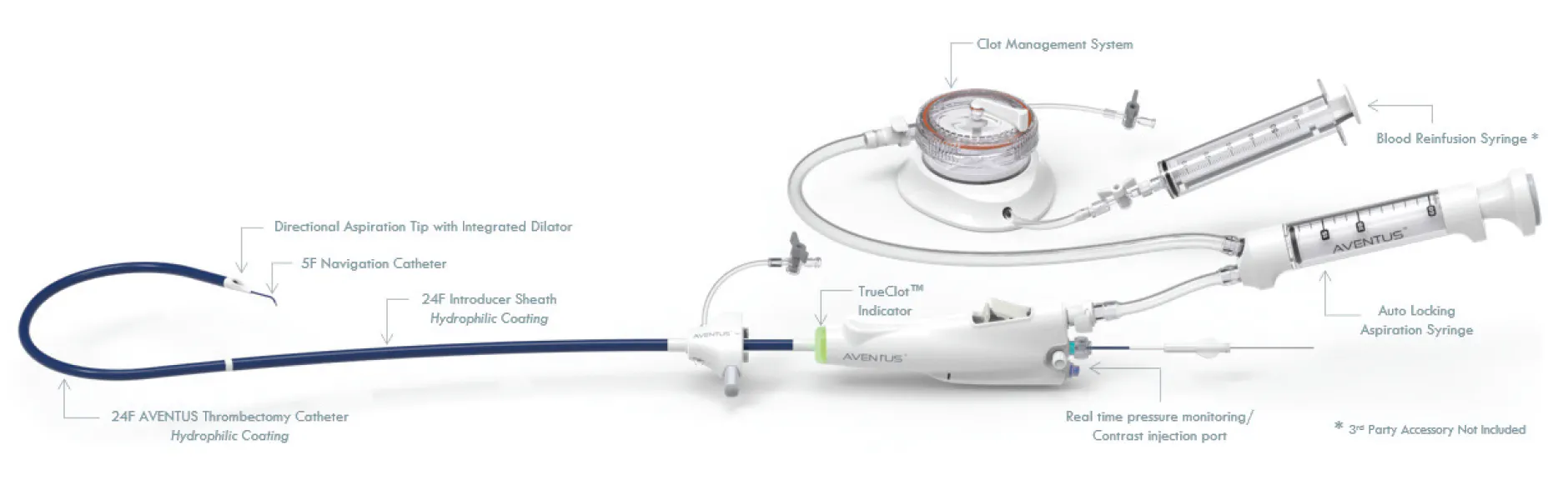
The AVENTUS® Thrombectomy System (Inquis Medical, Inc.) incorporates several integrated features designed to address these considerations: real-time tissue sensing at the catheter tip (TrueClot™ technology utilizing electrical impedance spectroscopy), an obliquely oriented aspiration tip enabling directional aspiration, and an inline clot canister that facilitates autologous blood return (Figure 1). This brief case series presents observations from a series of 40 consecutive cases, structured to highlight the procedural application of these three features.
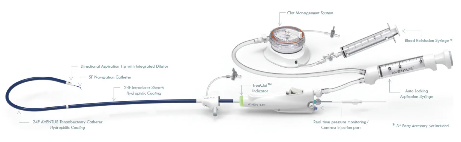
Figure 1. AVENTUS System overview.
CASE 1: REAL-TIME SENSING FOR THROMBUS DETECTION AND OPERATOR FEEDBACK
For our first case with the AVENTUS System, a male patient in his late 60s presented with intermediate-high–risk PE, including a right ventricular/left ventricular (RV/LV) ratio of 2.2:1, tachycardia (heart rate [HR], 128 bpm), and hypoxia (oxygen saturation, 82% on room air). CTA demonstrated bilateral large-volume lobar PE (Figure 2A).
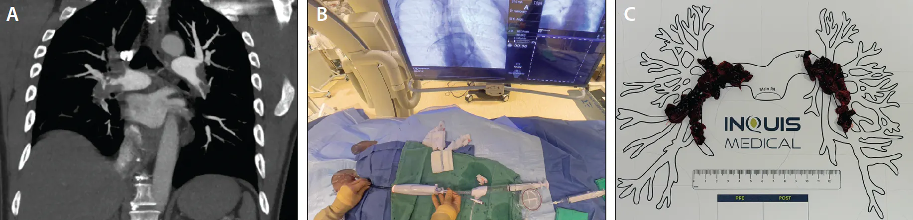
Figure 2. CTA of the chest showing large-volume bilateral PE (A). The sensing indicator on the AVENTUS Catheter handle illuminated orange, denoting interaction with thrombus at the catheter tip via real-time electrical impedance spectroscopy (TrueClot™ Sensing) (B). Large-volume thrombus, visually corresponding to the CTA images (C).
In our prior experience including several hundred large-bore aspiration thrombectomy cases, we would typically rely on angiographic landmarks that correspond to clot burden on CTA. This facilitates a thrombectomy technique in which the device is retracted during aspiration in a distal-to-proximal fashion.
Upon advancing the AVENTUS Catheter into the right PA, it tracked without resistance. The clot sensor indicator on the handle transitioned to orange sooner than anticipated, signaling engagement with thrombus in a more proximal location than expected based on angiographic landmarks (Figure 2B). Four aspirations were performed in this location, resulting in clearance of the right lobar thrombus burden. In additional targeted segments (such as lower lobe branches), the sensing indicator continued to provide confirmation of thrombus at the tip.
The immediate visual feedback from the sensing technology appeared to provide useful confirmation of tip-to-thrombus interaction beyond traditional landmarks. This correlation between the orange signal and productive aspiration cycles was observed to support operator confidence during catheter positioning—even on initial use—and may facilitate a shorter learning curve for achieving effective engagement, particularly for operators transitioning to this platform or large-bore PE thrombectomy in general. Clearance of targeted segments was accomplished efficiently with a limited number of aspiration cycles (Figure 2C).
This procedure was accomplished with eight aspirations in 11 minutes of device time, reducing mean PA pressure by 12 mm Hg. The patient was discharged home on postoperative day 2.
CASE 2: DIRECTIONAL ASPIRATION TO EASILY TARGET CLOT IN BRANCHES
A patient in their late 70s presented to the emergency department (ED) after a syncopal episode. He had mild tachycardia (108 bpm), bilateral segmental PE, a 2 L/min oxygen requirement, and an RV/LV ratio of 2.1:1. He was initially managed conservatively but experienced clinical deterioration over the subsequent 8 hours, with worsening oxygen requirements necessitating bilevel positive airway pressure, persistent tachycardia, and upward-trending serum troponin. The decision was made to proceed with MT.
After uneventful treatment of the right lower lobe (Figure 3A), the AVENTUS Catheter was advanced into the truncus anterior (TA). The beveled aspiration port was directed (via catheter torque) into the right upper lobe. This facilitated effective thrombus removal in a segment that can be challenging to engage fully with standard end-hole catheters.
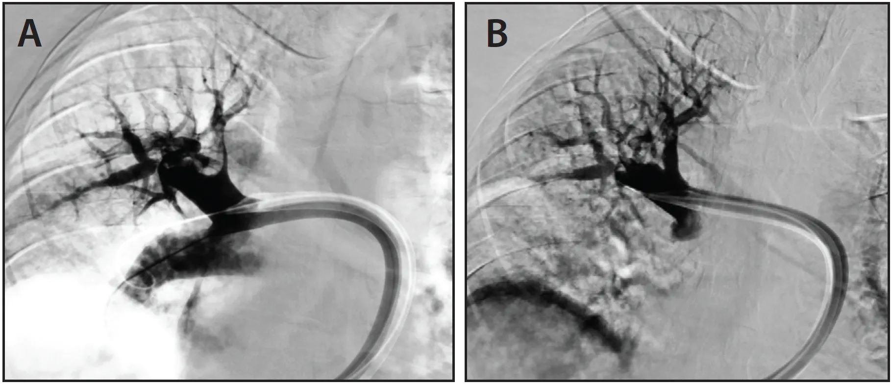
Figure 3. Filling defect in the apical segmental branch of the right upper lobe, reflecting occlusive segmental thrombus (A). Normal right upper lobe pulmonary angiogram after thrombectomy (B).
Effective thrombus removal from the targeted upper lobe segments was observed without requiring additional devices or repeated wire exchanges for redirection (Figure 3B).
Comparable utility of aspiration port orientation has been noted when addressing left lower lobe segmental disease in other procedures, where precise angling of the aspiration port facilitated engagement with clot located in anatomically challenging positions. The directional capability therefore appears particularly relevant in troubleshooting applications in locations such as the TA and left lower lobe branches.
CASE 3: AUTOLOGOUS BLOOD RETURN DURING HIGH-VOLUME ASPIRATION OF REFRACTORY THROMBUS
A male patient in his late 50s with a remote prior history of PE presented with a 2-week history of symptoms. On arrival to the ED, he was mildly tachycardic (HR, 103 bpm), required 6-L of oxygen via nasal cannula, remained normotensive, had an elevated serum troponin, and had an RV/LV ratio of 1.8:1. CTA demonstrated a large burden of bilateral PE (Figure 4A).
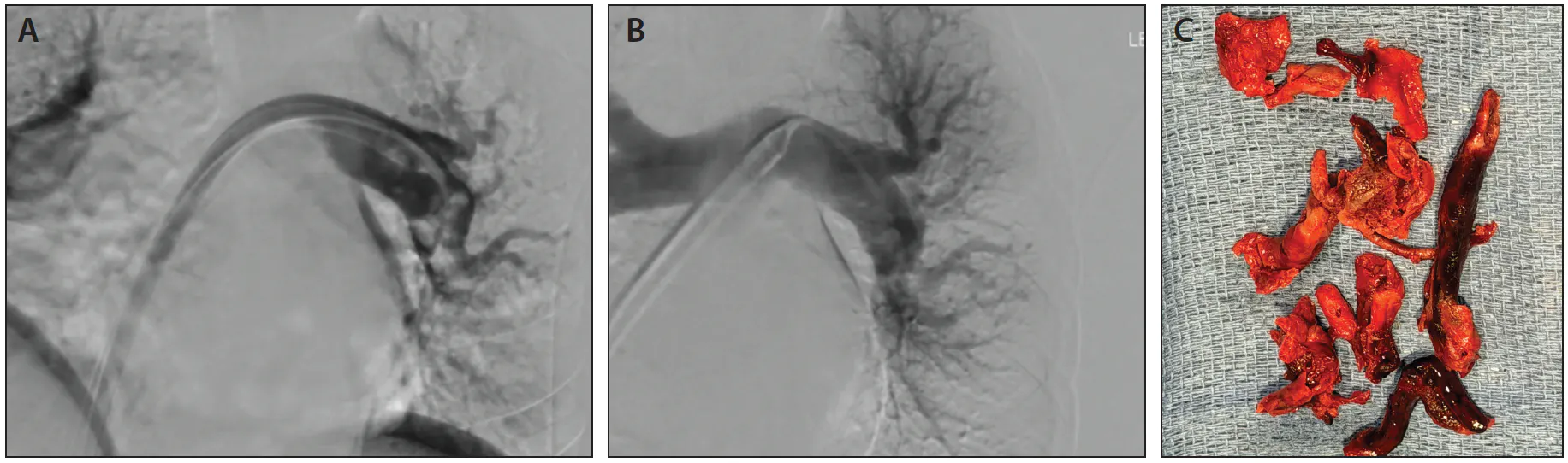
Figure 4. Pulmonary angiogram showing left lower lobe filling defects compatible with PE (A). Pulmonary angiogram following thrombectomy showing restoration of flow within the left lower lobe segmental PAs after numerous aspirations (B). Chronic, organized thrombus extracted with the AVENTUS System (C).
After successful thrombectomy of the lobar thrombus in the right PA, efforts to clear the left lower lobe were complicated by organized, chronic thrombus in a challenging medial segmental location. This required several aspirations over the course of 28 minutes of device time to fragment and remove the thrombus. The procedure was ultimately successful and resulted in normalization of pulmonary angiography (Figure 4B), large-volume thrombus removal, and 36 mm Hg reduction in systolic PA pressure.
After each aspiration into the syringe, the aspirated blood was quickly filtered through the AVENTUS Clot Management System. This system includes an integrated one-way valve connected directly from the aspiration catheter to the inline filtration chamber. Filtered blood was then returned to the patient while the thrombus was retained and visualized in the collection chamber. Despite a high number of aspiration attempts, net blood loss was limited to an estimated 120 mL. The removed material included substantial volumes of chronic, organized thrombus (Figure 4C).
This blood conservation feature proved especially valuable in this refractory scenario that demanded complex problem-solving and numerous aspirations. The case highlights the importance of effective autologous blood return in thrombectomy cases requiring extensive mechanical aspiration.
DISCUSSION AND OBSERVATIONS
These cases demonstrate the practical application of three design elements of the AVENTUS Thrombectomy System during PE intervention. The real-time tissue sensing provided continuous visual feedback at the handle that correlated with successful thrombus engagement and appeared to reduce nonproductive aspiration attempts while supporting operator positioning decisions. The obliquely oriented aspiration port enabled directional targeting in branched pulmonary anatomy, including the TA and left lower lobe segments, where conventional end-hole designs can be limited by port orientation relative to segmental clot. The integrated filtration and blood return system allowed maintenance of circulating volume, even when a large number of aspiration cycles were necessary for organized thrombus.
Navigation and aspiration were accomplished with a streamlined workflow that minimized device exchanges, consistent with the integrated dilator and navigation catheter design. Thrombus removal was achieved across a spectrum of clot chronicity, and no device-related complications (such as vessel injury or malfunction) were encountered in this early experience. The system handled both acute and more refractory thrombus burdens without apparent compromise in safety or efficiency.
Although these observations are preliminary and drawn from a limited series, the features appear to offer tangible procedural benefits in feedback, clot targeting precision, and blood management. The sensing technology in particular may assist with the adoption curve by supplying immediate, objective information about tip interaction with thrombus versus blood or vessel wall.
In a broader single-center experience encompassing > 40 cases at Huntsville Hospital in Huntsville, Alabama, the AVENTUS System was observed to aid procedural efficiency. Total device time averaged 14 minutes, including complete time for thrombectomy and blood reinfusion, resulting in minimal blood loss. Across these 40 cases, 89% of cases were completed with a single pass of the heart. Angiographic clot clearance was achieved in all cases, with no instances of technical failure or requirement for bailout devices.
Larger prospective series and comparative data will be required to quantify advantages in procedure time, blood loss, and clinical outcomes relative to other aspiration platforms.
CONCLUSION
The AVENTUS Thrombectomy System integrates real-time tissue sensing, directional aspiration capability via an obliquely oriented port, and autologous blood return into a single large-bore platform for PE thrombectomy. In this initial case experience, these elements were observed to function as described and to address specific practical challenges encountered during catheter-directed thrombus removal. The device demonstrated versatility across different anatomic locations and thrombus characteristics, with a safety profile consistent with expectations for contemporary aspiration thrombectomy. Continued clinical use and formal study data will help define its role in the management of PE.

Simplifying Large-Bore Venous Access: Introducing the AVENTUS™ Introducer Sheath
By Thomas Wong, MD
I am an interventional radiologist at Sarasota Memorial Hospital where we offer comprehensive, minimally invasive treatments for patients across Central Florida. One of our primary focus areas is venous thromboembolism (VTE) where we treat PE with access to most commercially available MT systems. While we are still in the early stages of evaluation, we have found that the AVENTUS® Thrombectomy System offers several unique features for VTE/PE treatment.
For our first AVENTUS cases, the AVENTUS™ Introducer Sheath (Inquis Medical, Inc.) was not yet available, so we utilized the Gore DrySeal 24-F sheath (Gore & Associates). Our team is skilled in using a variety of large-bore access sheaths, yet there are several considerations for the value of a purpose-built sheath for MT. When we heard that Inquis Medical was starting a limited market release of the new AVENTUS Sheath, we were excited to be one of the first hospitals in the United States to try it.
UNMET NEEDS IN LARGE-BORE VENOUS ACCESS
As MT continues to transform VTE/PE treatment, physicians are increasingly utilizing large-bore MT systems capable of rapidly removing significant thrombus burden. Although MT devices have advanced rapidly, large-bore venous access has remained an underappreciated technologic component.
Large-bore access introduces a unique set of clinical and workflow demands. Physicians must balance smooth delivery of increasingly large devices while maintaining hemostasis throughout the procedure. Most commercially available sheaths require dedicated focus or even two hands to manipulate valve pressure to control device insertion and advancement friction. Tedious valve adjustments can divert attention away from the patient, catheter, or guidewire position. Furthermore, MT requires a sheath to maintain a consistent hemostatic seal during transitions of devices from a 0.035-inch guidewire to a 24-F thrombectomy catheter.
Without continual focus on valve manipulation, physicians may encounter blood loss during exchanges, loss of guidewire position, or excess friction when manipulating thrombectomy catheters. Improving the ability to control hemostasis valve actuation allows the physician to maintain their focus on the procedure rather than the device.
Access site dilation is also an important component of venous access and device delivery. I typically start my MT procedures by taking a venogram and advancing a pigtail catheter to cross the heart, prior to dilating up to the 24-F sheath. Ultimately, the larger bore of the 24-F sheath requires a series of intermediate dilators. Therefore, especially in urgent PE cases where efficiency is paramount, the tapering of the introducer sheath can be critical in streamlining the time to first-pass intervention.
PE cases are demanding, and having an introducer sheath that is purpose-built to work seamlessly with MT systems is needed. To address these unmet needs, Inquis Medical, Inc. developed the AVENTUS Introducer Sheath, which is now commercially available.
AVENTUS INTRODUCER SHEATH OVERVIEW
The AVENTUS Introducer Sheath (Figure 1) was built specifically for the AVENTUS Thrombectomy System, combining innovative hemostasis technology with procedural control features designed to optimize every stage of access and MT catheter use.
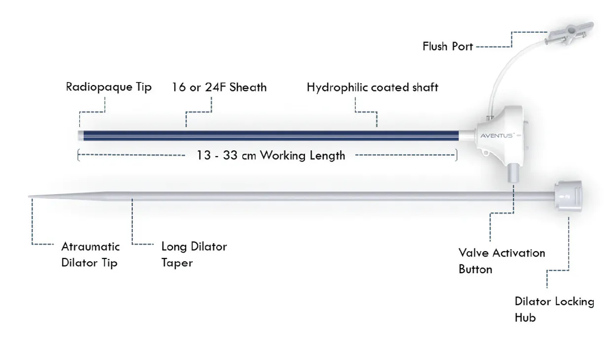
Figure 1. AVENTUS Introducer Sheath.
The AVENTUS Introducer Sheath utilizes dual-valve technology, and valve actuation is controlled by a single button that can be operated with a single hand.
Initial access is streamlined with a long taper dilator, a seamless dilator-to-sheath transition, and a hydrophilic sheath catheter. The result is noticeably smooth introduction into the access site and seamless control, dilating up to 24-F without the need for multiple intermediate dilators.
DUAL-VALVE TECHNOLOGY PROVIDING ADAPTIVE SEAL ACROSS A RANGE OF DEVICE PROFILES
At the core of the AVENTUS Sheath is an innovative dual-valve design that is engineered specifically for large-bore thrombectomy procedures.
A proximal iris-style valve (Figure 2) dynamically adapts to devices ranging from a 0.035-inch guidewire to a 24-F thrombectomy catheter, enabling hemostasis across a wide range of device profiles. Unlike traditional fixed-valve designs that require continual adjustments and manipulation while inserting devices, the proximal iris-style valve maintains constant hemostasis while reducing frictional forces against the catheter.
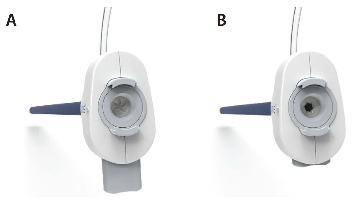
Figure 2. AVENTUS Introducer Sheath proximal valve in closed (A) and open (B) positions.
The valve conformity results in minimal resistance during dilator removal or manipulation of the AVENTUS Thrombectomy Catheter. Personally, this gives me confidence in maintaining guidewire position and streamlines procedural steps to stay focused on different steps in the thrombectomy procedure.
A secondary valve aids in ensuring hemostasis during fluid injections while maintaining a low drag force on any device inserted through the valve. The AVENTUS Introducer Sheath was designed to return blood with a fluid injection port and an adequate sheath inner diameter to provide clearance for injection. In my experience, blood reinfusion with the AVENTUS Introducer Sheath was noticeably smoother than other large-bore access sheaths we have used.
Together, the innovative design of the dual-valve system helps minimize blood loss, enables smooth device advancement, and optimizes procedural efficiency.
SIMPLIFIED VALVE CONTROL WITH A SINGLE ACTUATION BUTTON
The AVENTUS Sheath incorporates an intuitive single-button valve actuation mechanism designed for efficient one-handed operation (Figure 3). The actuation mechanism allows physicians to easily titrate the valve opening during device insertion, while maintaining hemostasis. Once the AVENTUS Thrombectomy Catheter is inserted past the valve, the actuation button no longer needs to be depressed.
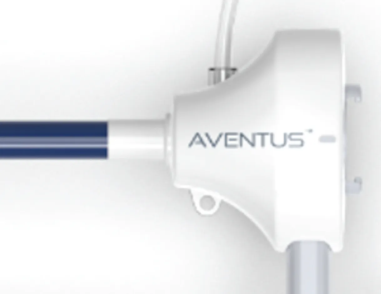
Figure 3. AVENTUS Introducer Sheath with single button and single-handed valve actuation.
Having a sheath that is simple to control and allows for free manipulation of inserted devices enables me to focus on other aspects of the procedure. By reducing complexity at the access site, the AVENTUS Introducer Sheath supports faster device insertion and smoother exchanges throughout the procedure.
ENGINEERED FOR SMOOTH VENOUS ACCESS AND DILATATION
The AVENTUS Introducer Sheath was designed to facilitate controlled insertion and advancement during large-bore venous access procedures.
The dilator has a small-caliber tip and a longer taper compared to other large-bore sheaths we have used. The dilator-to-sheath transition is incredibly smooth, and the sheath catheter has a hydrophilic coating. These elements translate into noticeably smooth access site insertion and the ability to dilate up to 24-F without reliance on intermediate dilators. In urgent PE cases, this means less time before our first aspiration is completed or less time before we get the patient’s hemodynamics stabilized.
Removal of the dilator is simple, requiring only a 90° twist to unlock from the sheath. Removing the dilator is very smooth, which is important to help maintain both sheath and guidewire positioning.
The sheath catheter has a higher kink resistance than other large-bore sheaths, resulting in a more flexible and trackable access sheath. I have found this beneficial, as our PE patients are not under heavy sedation and sometimes they will complain of lower back discomfort with sheaths that are too rigid. Femoral access and insertion into the inferior vena cava (IVC) are not overly tortuous, but there is some angulation, and a flexible sheath catheter improves patient comfort and tolerance of the whole procedure.
Collectively, these design elements improve large-bore venous access and support rapid thrombectomy intervention.
PURPOSE-BUILT FOR THE NEXT GENERATION OF MT
As large-bore thrombectomy procedures continue to evolve, access site management is a critical aspect of overall procedural performance. The new AVENTUS Introducer Sheath offers several favorable advantages and it pairs extremely well with the AVENTUS Thrombectomy System. The AVENTUS Sheath is purpose-built to address the practical challenges physicians face during MT, with simplified valve operation, controlled hemostasis, and easy access.
In my experience, the AVENTUS Thrombectomy System offers several features that may advance the field of MT. Pairing the AVENTUS Introducer Sheath with the AVENTUS Thrombectomy System further streamlines treatment for VTE and PE.

CASE REPORT
Step-by-Step Management of PE With the AVENTUS® Thrombectomy System
By Ripal T. Gandhi, MD, FSVM, FSIR
VTE affects more than 900,000 individuals annually in the United States, with acute PE representing the third-leading cause of cardiovascular mortality. Despite standard anticoagulation (AC) therapy, the risks of morbidity and mortality remain substantial. In addition to its acute clinical impact, PE is associated with significant long-term impairment in quality of life due to post-PE syndrome and chronic thromboembolic pulmonary hypertension (CTEPH).
A meta-analysis of nearly 3,700 patients with PE and a median follow-up of 18 months demonstrated persistent RV dysfunction in 18% of patients and moderate-to-severe functional limitation (New York Heart Association class III-IV) in 11%.1 Affected patients exhibited markedly reduced exercise capacity, with 6-minute walk test performance at approximately the 5th percentile. Early and effective thrombus removal in the setting of acute PE may reduce the risk of PE-related mortality, clinical deterioration, cardiorespiratory collapse, recurrent embolic events, post-PE syndrome, and the development of CTEPH.
This case highlights my step-by-step approach to the management of acute PE using the AVENTUS® Thrombectomy System, with an emphasis on practical clinical insights, technical considerations, and procedural strategies to address real-world challenges encountered during MT.
CASE PRESENTATION
A male patient in his early 60s with a medical history significant for hypertension and prior embolization of a dural venous fistula presented with a 2-week history of cough, chest pain, and progressively worsening dyspnea.
On presentation, he was normotensive with a blood pressure (BP) of 126/91 mm Hg and tachycardic with a baseline HR of 108 bpm. Respiratory rate was 22 breaths/minute and oxygen saturation was 90% on 2 L of supplemental oxygen. With minimal exertion, including ambulation to the bathroom, his HR increased to the 130s bpm and oxygen saturation decreased to 87%, accompanied by marked dyspnea.
Laboratory evaluation demonstrated elevated cardiac and hemodynamic biomarkers, including an NT-proBNP of 1,461 pg/mL, troponin of 1,188 ng/L, and lactic acid of 2.4 mmol/L. CT pulmonary angiography (CTPA) revealed extensive bilateral PE involving the bilateral PAs, with evidence of RV strain demonstrated by an RV/LV ratio of 1.6 (Figure 1 and Figure 2). Transthoracic echocardiography (TTE) demonstrated a dilated right ventricle with moderately reduced systolic function, as well as pulmonary hypertension with an estimated RV systolic pressure of 40 mm Hg (Figure 3).
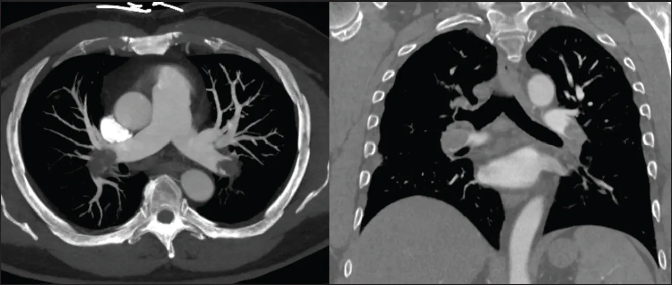
Figure 1. CTPA demonstrating significant PE involving the bilateral PAs.
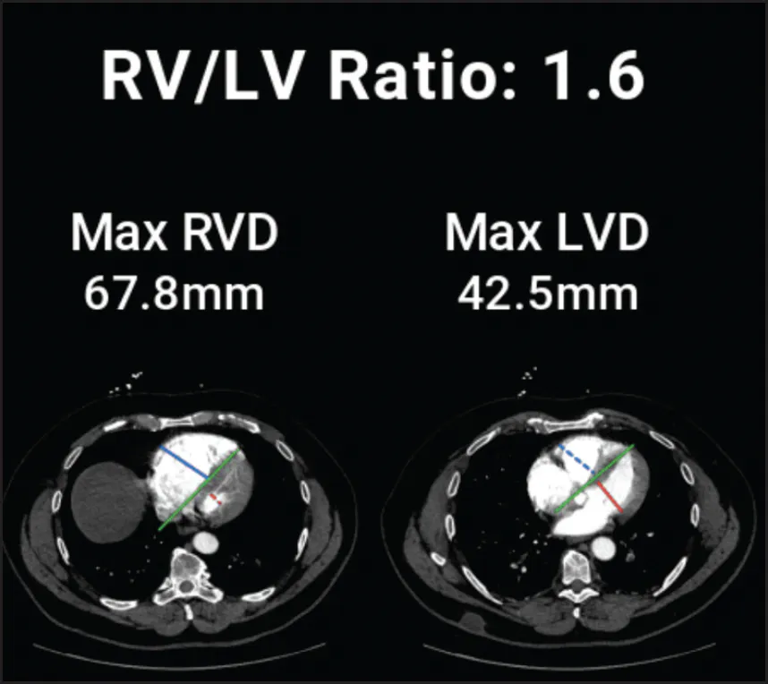
Figure 2. Artificial intelligence technology (Viz.ai) demonstrating an elevated RV/LV ratio of 1.6.
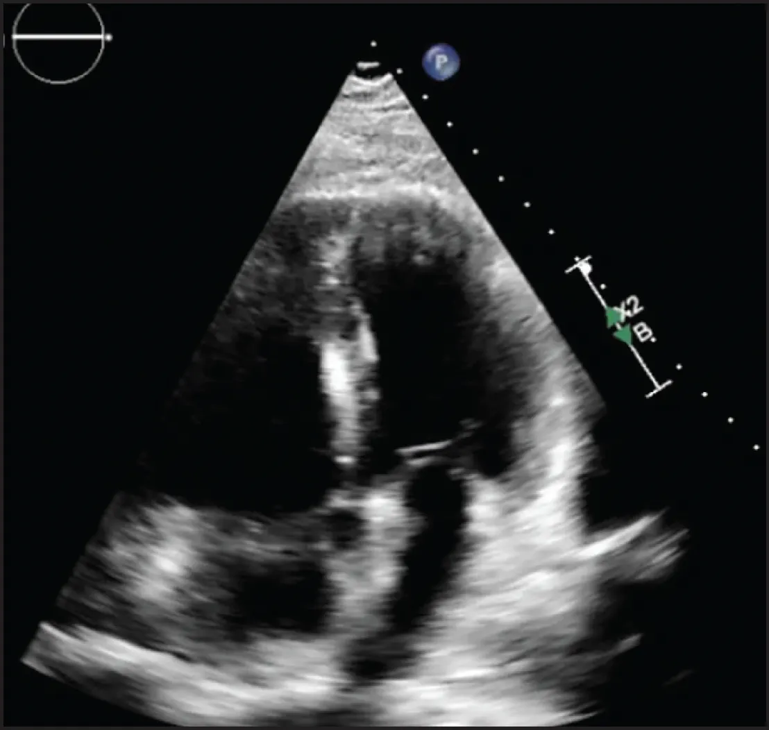
Figure 3. TTE demonstrated a dilated right ventricle with moderately reduced systolic function. Estimated RV systolic pressure was 40 mm Hg.
RATIONALE FOR INTERVENTION
According to the 2026 American Heart Association/American College of Cardiology (AHA/ACC) guidelines for the management of acute PE in adults, the patient met criteria for incipient cardiopulmonary failure with normotensive shock, based on the presence of elevated lactic acid, corresponding to an AHA/ACC acute PE clinical category D2.2 Notably, with minimal exertion, the patient’s shock index (HR/systolic BP) increased to 1.03, raising concern for significant hemodynamic stress and evolving shock physiology. The updated guidelines provide a class IIb recommendation for this patient population, stating that in patients with acute PE classified as AHA/ACC PE categories D1 to D2 in whom advanced therapy is being considered, MT in addition to AC may be considered over AC alone to reduce the risk of further clinical deterioration.
Our institutional philosophy and approach is to enroll all eligible patients in clinical trials whenever feasible; however, the patient declined participation. Accordingly, the decision was made to proceed with MT using the AVENTUS Thrombectomy System.
PROCEDURAL OVERVIEW
Ultrasound-guided venous access was obtained via the right common femoral vein, and venography of the IVC and right iliac venous system was performed to exclude the presence of thrombus. Using an angled pigtail catheter, the right heart was traversed and the right PA was selectively engaged. Hemodynamic assessment demonstrated markedly elevated PA pressures of 84/28 mm Hg (mean, 49 mm Hg). Pulmonary angiography revealed a large thrombus burden within the right PA and TA (Figure 4).
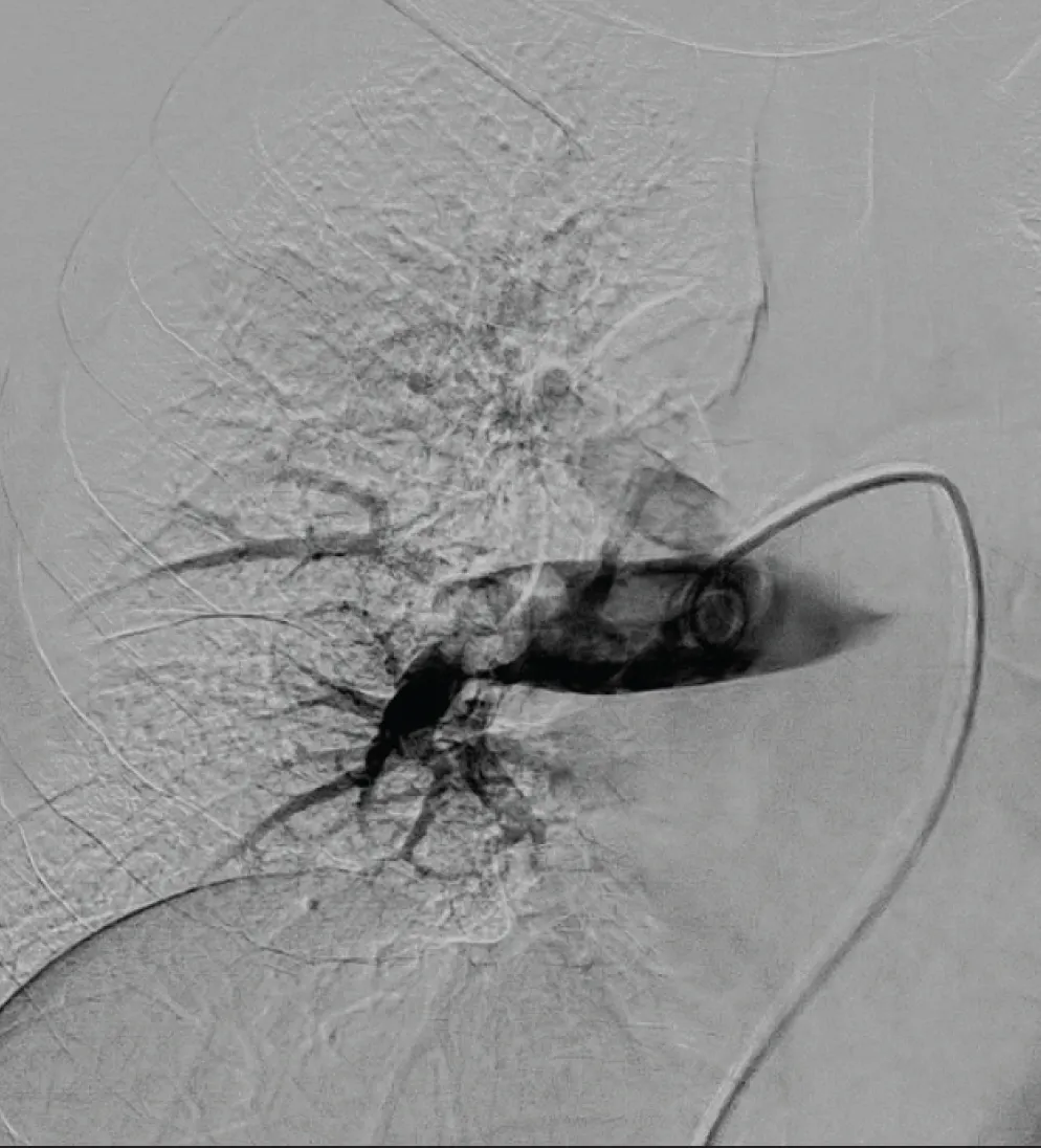
Figure 4. Large PE involving the right main PA and TA.
The AVENTUS Thrombectomy System was advanced over a 1-cm floppy-tip Amplatz guidewire into the right PA (Figure 5). MT was then performed with controlled rotation of the device to optimize thrombus targeting and extraction. The directional aspiration tip was oriented superiorly to facilitate aspiration of thrombus from the TA. The directional aspiration tip design facilitates effective thrombus extraction without the need for selective cannulation of each individual PA branch.
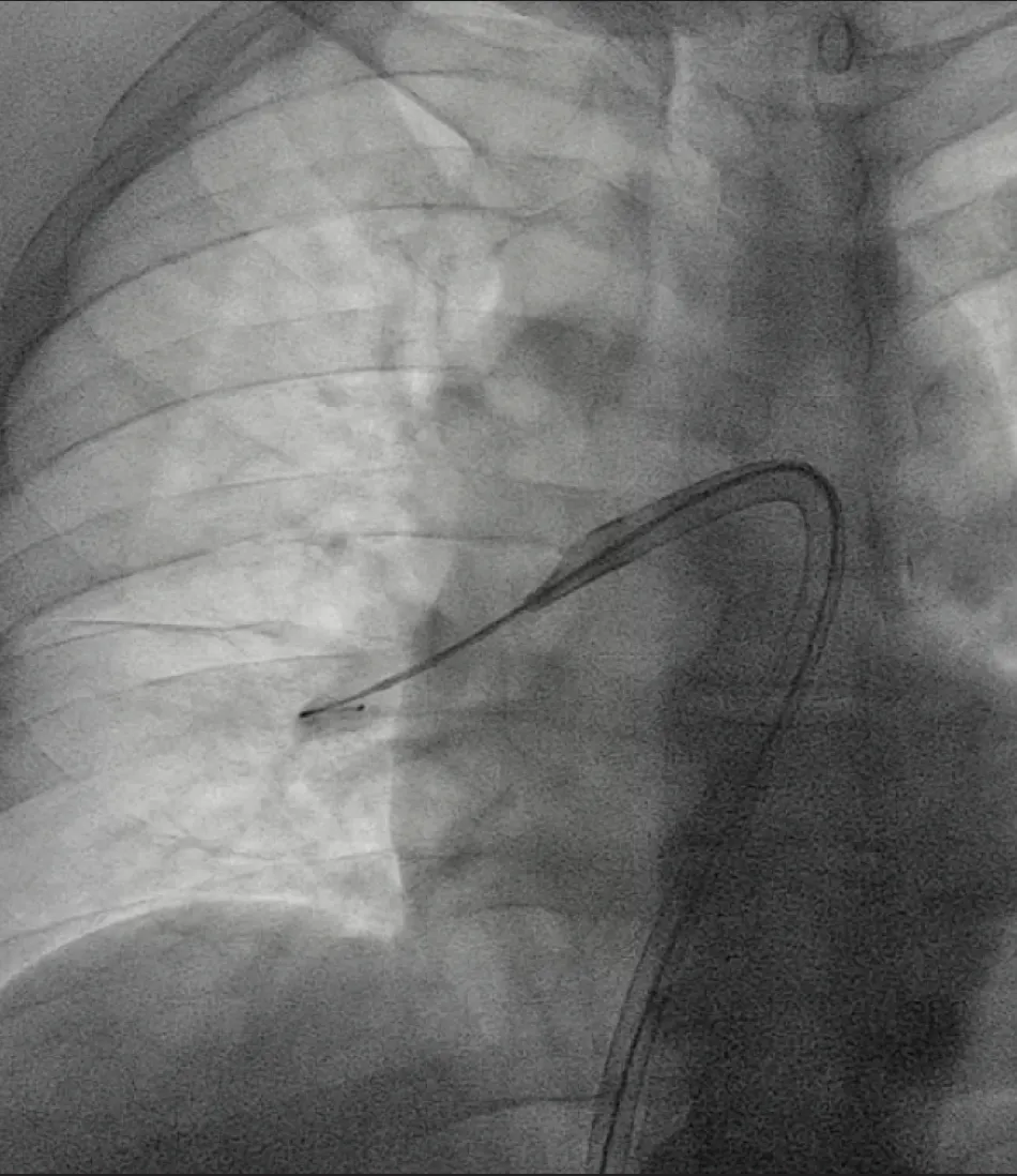
Figure 5. AVENTUS System positioned within the right PA. The device may be rotated as needed to facilitate targeted thrombus extraction from multiple PA branches. The directional catheter tip is oriented superiorly to engage thrombus within the TA.
The AVENTUS System incorporates TrueClot™ Sensing technology designed to enhance procedural efficiency during thrombectomy. The system provides real-time feedback, with green indicating free-flowing blood, orange indicating thrombus engagement, and blue indicating vessel wall apposition. Figure 6 shows the orange indicator, consistent with active thrombus engagement. This information equips the operator with real-time feedback of what the catheter tip is in contact with, providing information to help complete the procedure faster.
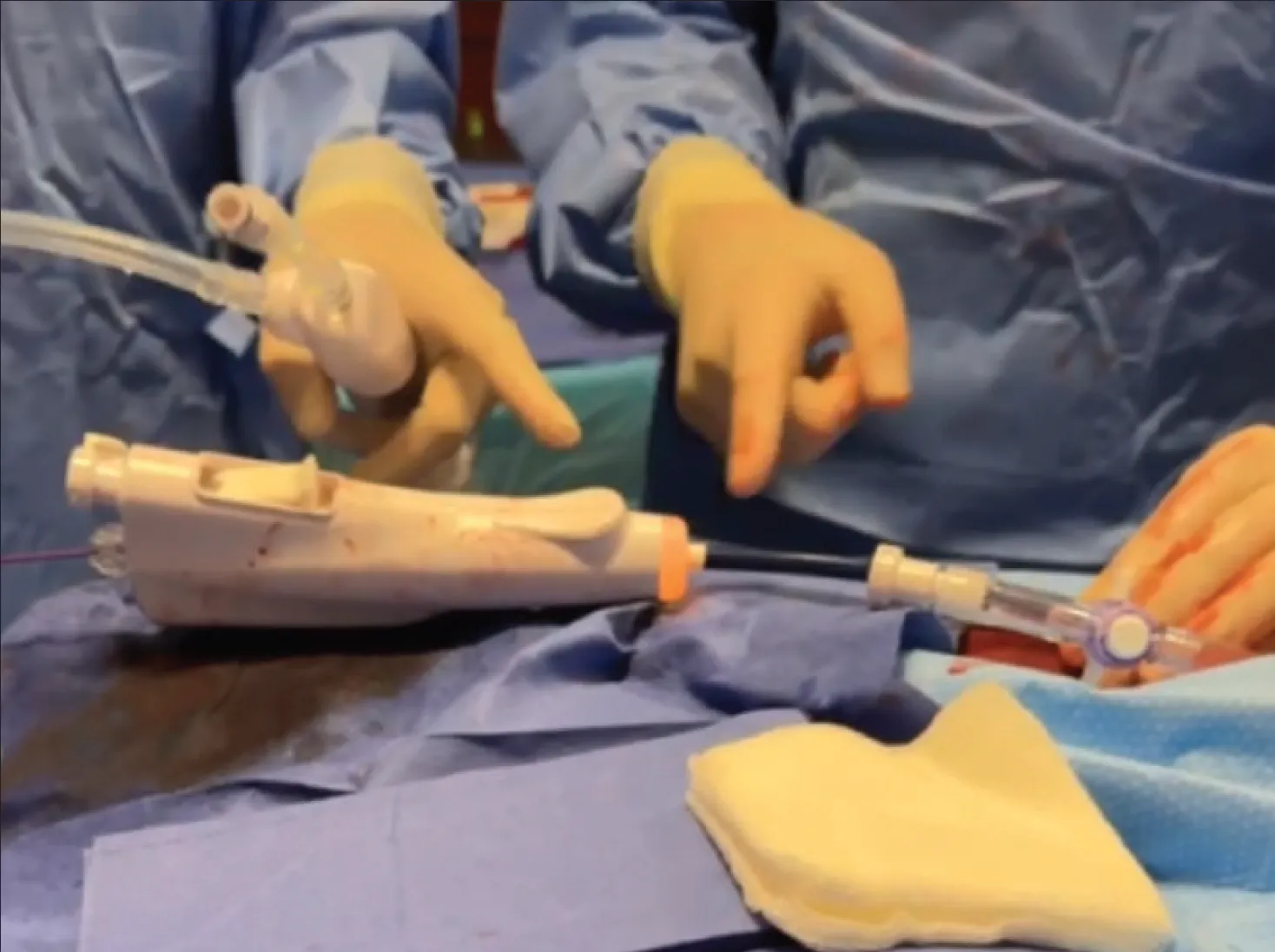
Figure 6. The orange indicator denotes detection of thrombus at the catheter tip, indicating optimal positioning for active aspiration.
Following thrombus aspiration, blood reinfusion was performed via the in-line reinfusion system, which is designed to reduce manual steps and thereby improve procedural efficiency and shorten overall procedure time (Figure 7). The aspiration syringe remains continuously connected to the catheter throughout the procedure, thereby reducing procedural steps and eliminating the need for syringe detachment. This results in fast and clean clot visualization, blood filtration, and blood return all at the patient table in front of you.
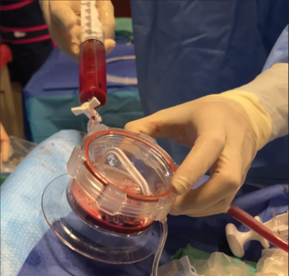
Figure 7. Streamlined blood reinfusion using an in-line filtration and reinfusion system. After each aspiration cycle, the syringe is advanced, directing blood and thrombus into the clot canister via a dual one-way valve system. Within the canister, dual filtration mechanisms separate thrombus from blood, enabling immediate visualization of extracted material and real-time confirmation of aspiration efficacy.
Final right pulmonary angiography demonstrated successful removal of the majority of the thrombus burden (Figure 8).
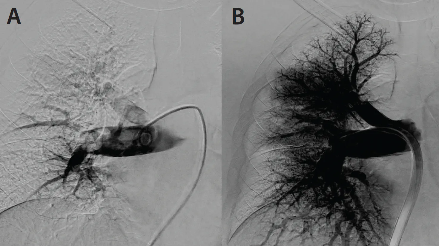
Figure 8. Pre- (A) and post-thrombectomy (B) imaging of the right PA demonstrating significant improvement in PA flow with minimal residual thrombus.
The AVENTUS Thrombectomy System was easily transitioned from the right to the left PA through simple retraction of the device and guidewire, allowing the system to naturally redirect into the left PA. The system includes a 5-F navigation catheter, which facilitates efficient guidewire advancement into the target location and subsequent device delivery without the need for a dilator. Eliminating the need for a dilator when advancing the device contributes to meaningful procedural time savings.
The directional aspiration was then rotated inferiorly to facilitate successful aspiration of thrombus within the lower left PA (Figure 9).
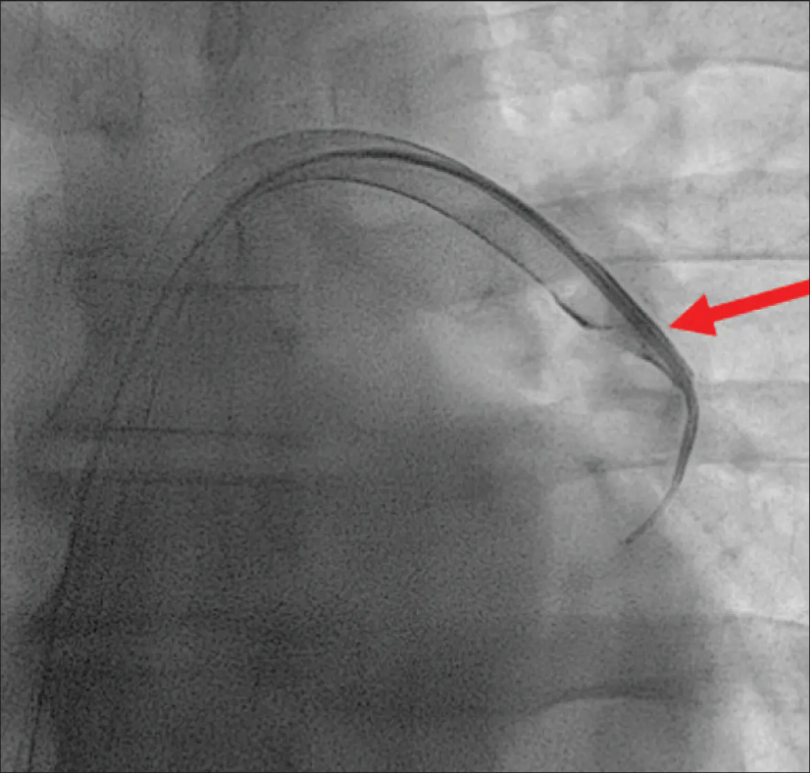
Figure 9. AVENTUS System with directional aspiration positioned caudally to remove thrombus from the left PA.
Final left pulmonary angiogram demonstrated successful removal of the majority of the left-sided PE, with small residual distal thrombus (Figure 10). The patient became increasingly restless, and the procedure was subsequently concluded, precluding further intervention on the residual thrombus.
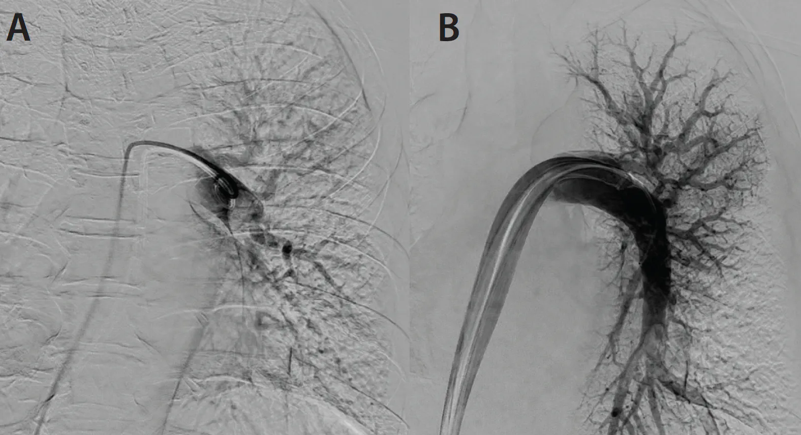
Figure 10. Pre- (A) and post-thrombectomy (B) images of the left PA.
Final PA pressures after thrombectomy were 38/18 mm Hg (mean, 22 mm Hg), representing a 46 mm Hg reduction in systolic PA pressure. Figure 11 illustrates the extracted thrombus.
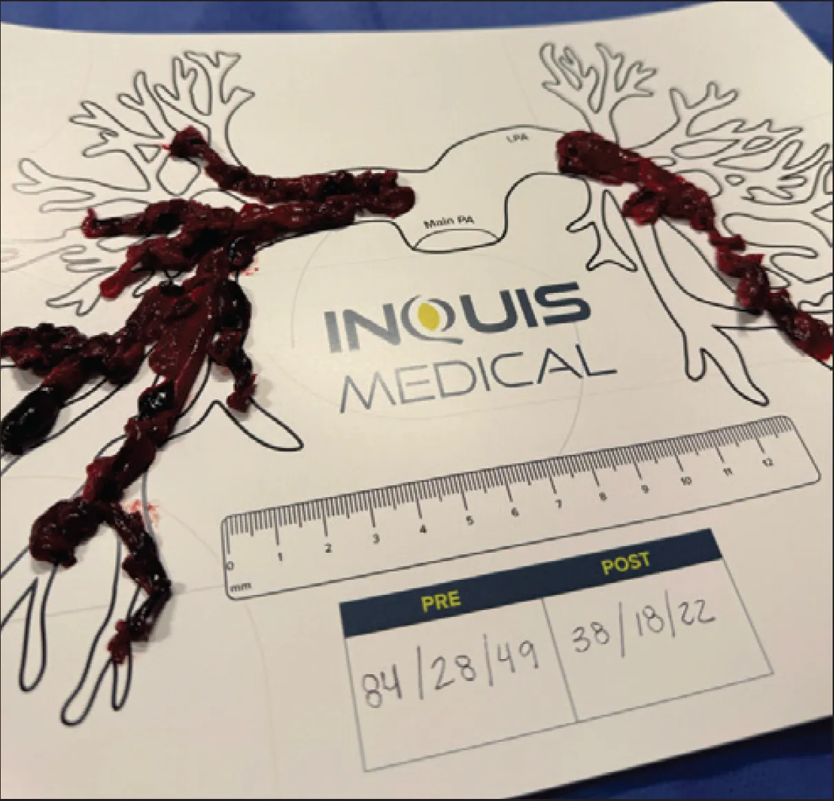
Figure 11. Thrombus extracted during the procedure. Note the significant drop in PA pressures.
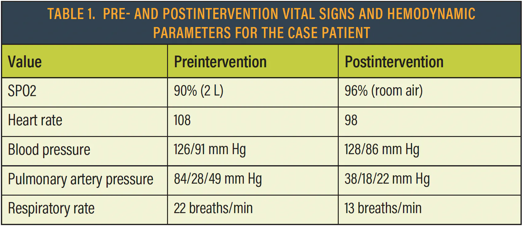
Most importantly, the patient experienced complete resolution of symptoms. A postprocedural ambulation test performed on postoperative day one demonstrated an oxygen saturation of 96% with an HR of 98 bpm. Table 1 compares pre- and postintervention vital signs and hemodynamic parameters, demonstrating marked improvement across all measured variables.
POSTINTERVENTION RESULTS
A TTE obtained 24 hours postprocedure demonstrated normalization of RV size, morphology, and wall thickness, with restoration of normal RV systolic function. In addition, repeat CTPA performed 48 hours postprocedure demonstrated near-complete resolution of the thrombus burden (Figure 12).
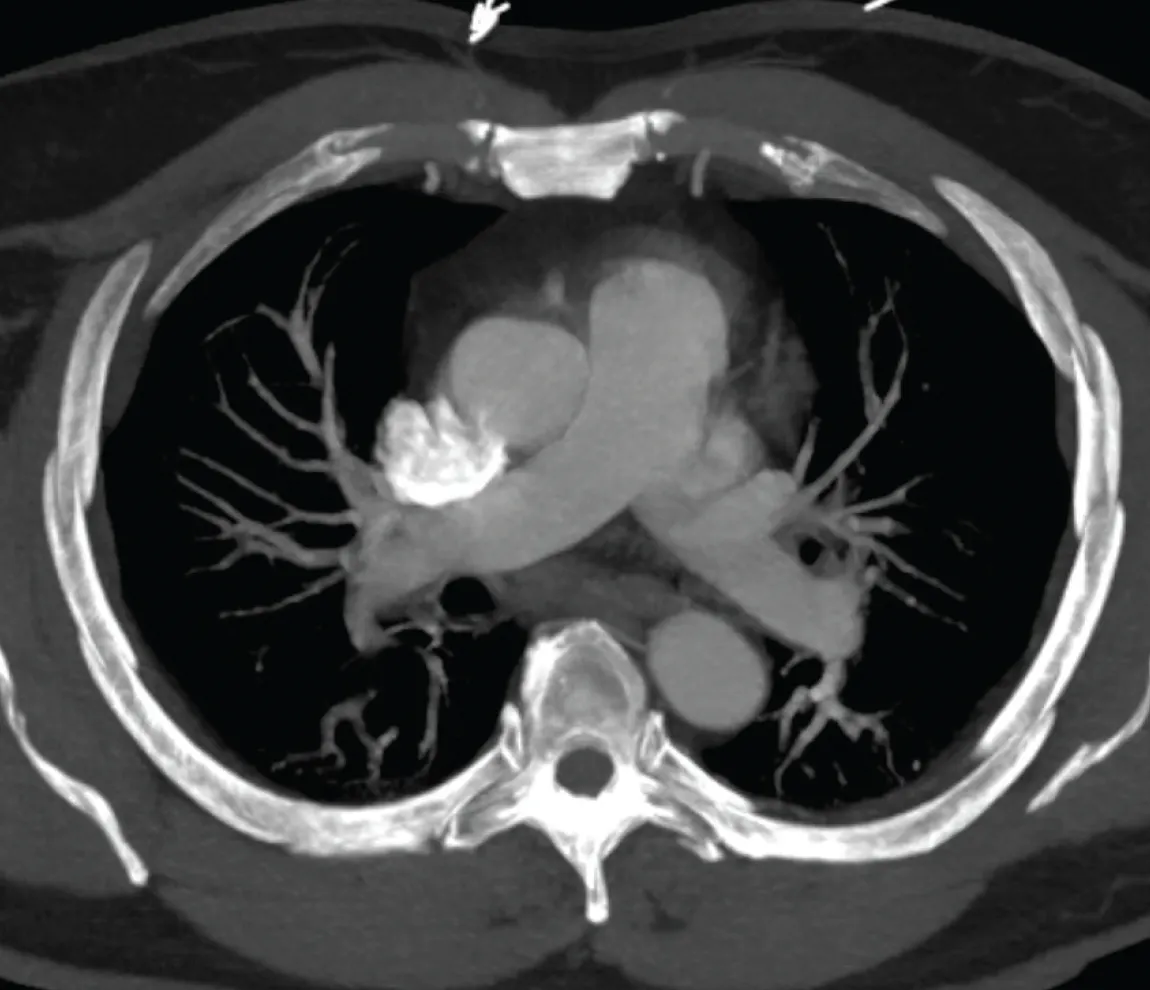
Figure 12. CTPA at 48 hours post-treatment demonstrating minimal residual thrombus burden.
Total blood loss was 30 mL, reflecting efficient aspiration and effective blood reinfusion. Only a single passage of the device through the right heart was required, with a total of two aspiration cycles performed (one in each PA), both corresponding to an orange sensor reading indicative of thrombus engagement.
DISCUSSION
The AVENTUS Thrombectomy System represents an emerging advancement in the percutaneous treatment of PE, enabling efficient thrombus extraction with concurrent blood reinfusion, while enhancing procedural safety and precision through TrueClot™ Sensing technology.
The case demonstrates the advantage of the directional aspiration tip, allowing the device to be rotated to extract thrombus from multiple branches without the need for selective branch cannulation, resulting in meaningful time savings. The entire right PA, including the TA, was successfully treated with a single aspiration by orienting the directional aspiration tip cranially during slow catheter retraction (Figure 13). Treatment of the left PA can be particularly challenging with other end-hole MT devices due to its acute angulation and posterior orientation, which increase the likelihood of vessel wall latch. The AVENTUS Thrombectomy System is specifically designed to facilitate intervention in the left PA by directing the catheter toward the thrombus while avoiding contact with the arterial wall (Figure 13). In this case, the entire left PA was also successfully treated with a single aspiration.
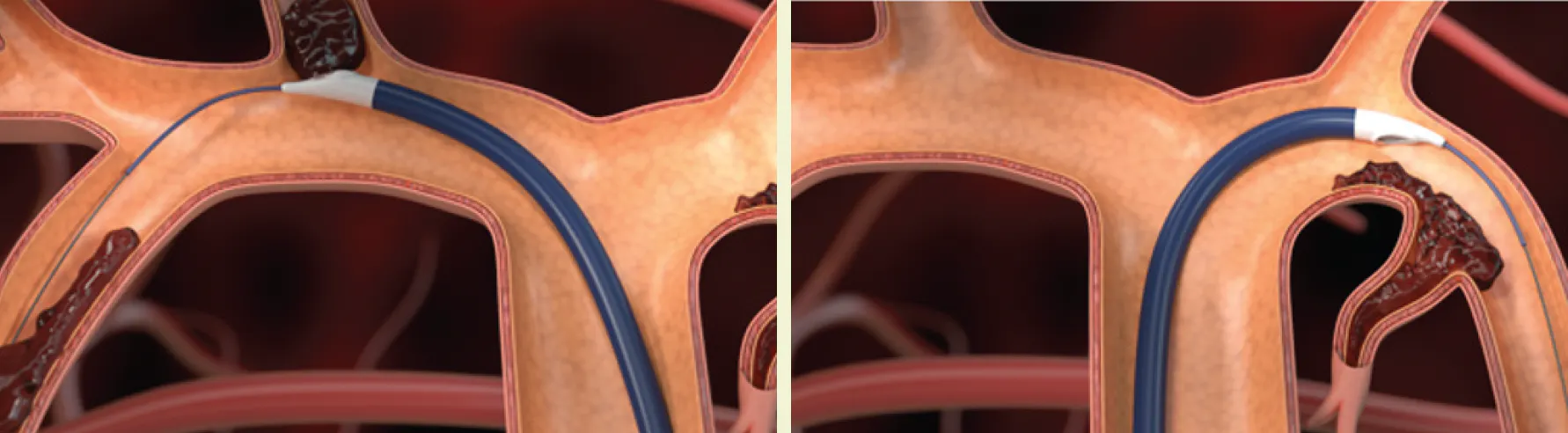
Figure 13. The catheter’s directional aspiration technology enhances procedural efficiency. It allows the operator to direct aspiration toward the thrombus without having to wire each individual branch. Directional aspiration also enables clot removal from segmental branches while remaining in the main PA, without repositioning the guidewire or selectively catheterizing each branch.
Another advantage of the directional aspiration tip is that its cross-sectional area is larger than that of comparable end-hole devices of the same French size. In addition, the angled configuration leverages fluid dynamics to create a vortex effect, enhancing clot retrieval compared with end-hole designs.
Proprietary TrueClot™ Sensing technology provides real-time feedback on conditions at the catheter tip, including free-flowing blood, wall latch, lollipop thrombus, and catheter clogging. This capability improves procedural efficiency by reducing empty aspirations and allowing for real-time troubleshooting, precluding unnecessary removal of the catheter from the body.
The AVENTUS Thrombectomy System is one of only two systems that are FDA cleared for blood reinfusion. It features a streamlined in-line reinfusion process performed at the table within a closed, controlled system, which minimizes blood loss, reduces procedural steps, and shortens overall procedure time. The importance of blood reinfusion cannot be overstated as studies have shown that blood return is associated with lower mortality and decreased need for blood transfusion.3
Other advantageous features of the device include the ability to monitor PA pressures in real time, excellent trackability and torque response, and a tapered design that eliminates the need for a dilator. Additional benefits include minimal guidewire exchanges and the ability to advance the device over a softer guidewire. Finally, the system has a very short learning curve, with operator familiarity achievable very quickly. In fact, I was comfortable using the device after a single case.
The AVENTUS Thrombectomy System represents a next-generation approach to MT for PE, incorporating innovative features that enhance clinical efficiency and reduce procedural time while maintaining safety.
1. Sista AK, Miller LE, Kahn SR, Kline JA. Persistent right ventricular dysfunction, functional capacity limitation, exercise intolerance, and quality of life impairment following pulmonary embolism: systematic review with meta-analysis. Vasc Med. 2017;22:37-43. doi: 10.1177/1358863X16670250
2. Creager MA, Barnes GD, Giri J, et al. 2026 AHA/ACC/ACCP/ACEP/CHEST/SCAI/SHM/SIR/SVM/SVN guideline for the evaluation and management of acute pulmonary embolism in adults: a report of the American College of Cardiology/American Heart Association Joint Committee on Clinical Practice Guidelines. Circulation. 2026;153:e977-e1051. doi: 10.1161/CIR.0000000000001415
3. Ali B, Patel D, Arora S, et al. Impact of a blood return system on mechanical thrombectomy in treatment of acute pulmonary embolism: a retrospective cohort study comparing 30-day mortality and need for blood transfusion. Eur Heart J. 2024;45(suppl 1):ehae666.2186. https://doi.org/10.1093/eurheartj/ehae666.2186

CASE REPORT
Fast and Precise Clot Removal in Intermediate-High–Risk PE Using the AVENTUS® Thrombectomy System
By Patrick Muck, MD
PE remains one of the most challenging acute cardiovascular conditions encountered in modern medicine. Although advances in diagnostic imaging, risk stratification, and catheter-based therapies have expanded treatment options, optimal outcomes still depend on the ability to rapidly identify high-risk patients and coordinate care across multiple specialties. At TriHealth in Cincinnati, Ohio, this challenge is addressed through a well-established PE response team that brings together vascular surgery, interventional cardiology, pulmonary critical care, emergency medicine, hematology, cardiothoracic surgery, and advanced practice providers to deliver timely, patient-centered care.
Our approach begins with a comprehensive assessment of hemodynamic status, RV function, biomarker elevation, clot burden, bleeding risk, and overall patient condition. Although AC remains the foundation of therapy for many patients, those presenting with intermediate-high–risk PE and evidence of RV strain may benefit from MT to rapidly reduce clot burden, improve cardiopulmonary function, and potentially prevent clinical deterioration.
As MT technology continues to advance, there is increasing emphasis not only on effective clot removal but also on procedural efficiency and the ability to address thrombus located in branch PAs. Directional aspiration technologies have introduced new opportunities to precisely target residual clot while minimizing the need for additional catheter manipulation or guidewire repositioning.
This case highlights the treatment of a young female patient who presented with intermediate-high–risk PE and significant RV strain. The case demonstrates how the directional aspiration capabilities of the AVENTUS® Thrombectomy System facilitated efficient thrombus removal from both main PA branches and the TA without requiring repositioning of the guidewire, resulting in immediate clinical improvement and restoration of pulmonary blood flow.
CASE PRESENTATION
A female patient in her mid 30s presented to the ED with progressive shortness of breath and chest pain that had been worsening over the previous week. Upon arrival, she was tachycardic and hypertensive. CTA demonstrated bilateral PE with significant right heart strain, including an RV/LV ratio of 1.6. Coronal and axial CTA showed bilateral clot distribution, with occlusion of both the upper and lower lobes of the right and left PA (Figure 1 and Figure 2). The TA of the right PA also appeared to be fully occluded. Laboratory testing revealed elevated cardiac biomarkers, with a troponin of 58 ng/L and BNP of 176 pg/mL. Blood pressure was 129/88 mm Hg. On admission, the patient weighed 202 lb and height was 66 inches.
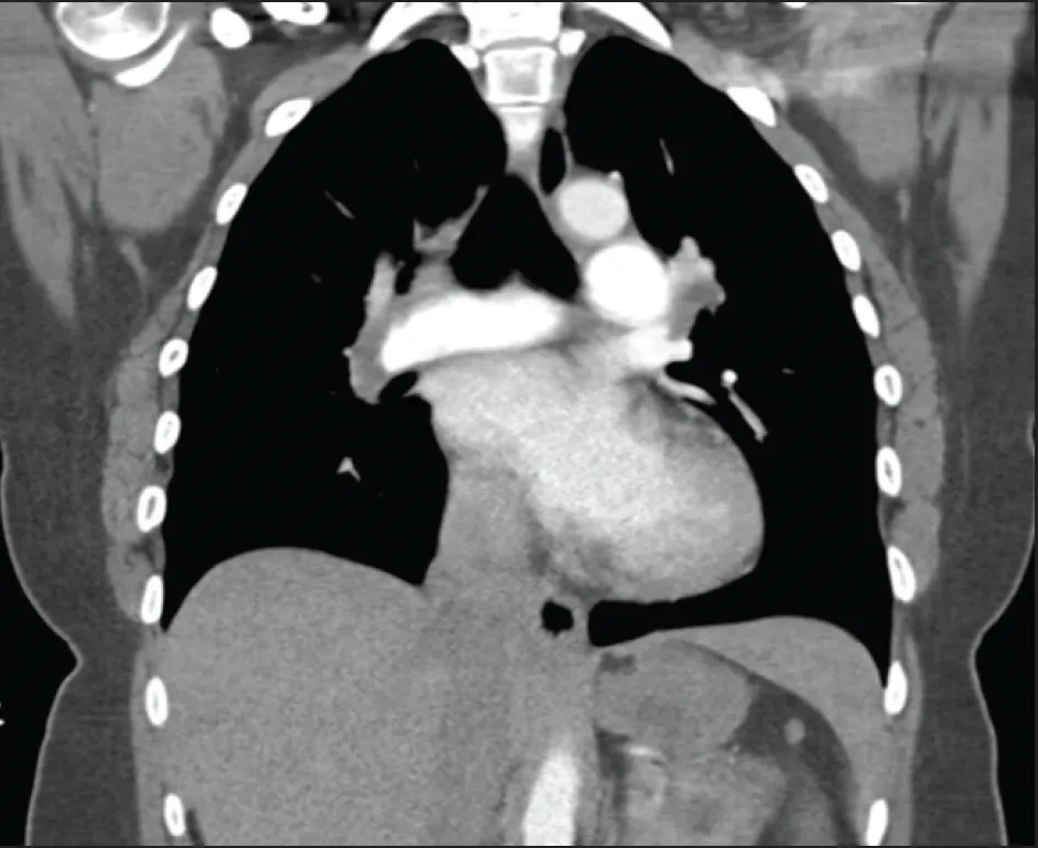
Figure 1. Coronal CTA showing clot present in both the left and right PA.
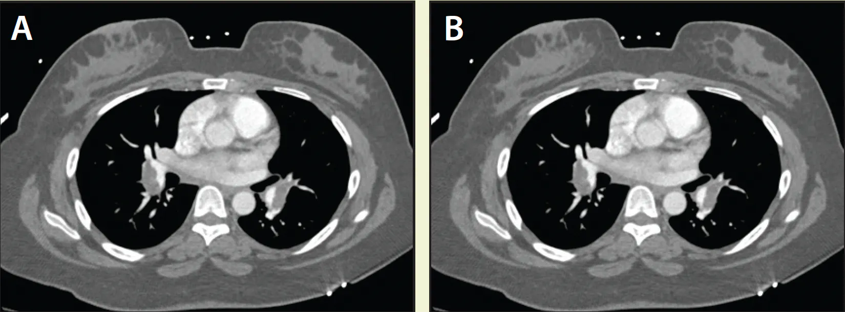
Figure 2. Axial CTA confirming clot present in both the upper and lower branches of the right (A) and left PA (B).
The patient’s medical history was notable for the absence of prior VTE, recent surgery, prolonged travel, COVID-19 infection, malignancy, or family history of hypercoagulability. Further investigation identified initiation of oral contraceptive therapy 3 weeks prior for treatment of heavy menstrual bleeding. The patient discontinued the medication after developing significant dyspnea.
TTE demonstrated preserved LV function with an estimated ejection fraction of 55% to 60%, mild concentric LV hypertrophy, mild tricuspid regurgitation, and mildly elevated RV systolic pressure of 35 to 45 mm Hg. Additional findings included right atrial dilation, interventricular septal flattening consistent with RV pressure overload, and moderately reduced RV systolic function.
Based on the imaging findings, biomarker elevation, and evidence of RV dysfunction, the patient was diagnosed with intermediate-high–risk PE.
PROCEDURAL OVERVIEW
Given the significant clot burden and evidence of right heart strain, the decision was made to proceed with MT utilizing the AVENTUS Thrombectomy System. With clot occluding several branches of the PAs, we felt directional aspiration could help in targeting multiple branches.
Ultrasound-guided access was obtained in the right common femoral vein and initially upsized to an 8-F sheath. An angled pigtail catheter was advanced into the PA, traversing the tricuspid valve safely without interaction with the chordae tendineae. Bilateral pulmonary angiography confirmed extensive thrombus burden in both the left and right PAs (Figure 3).
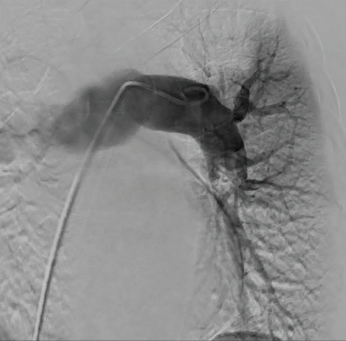
Figure 3. Angiography of the left PA showing occlusion of both the upper and lower lobes
After angiographic assessment, the 8-F sheath was exchanged for a Gore DrySeal 24-F sheath, and MT was performed using the AVENTUS Thrombectomy System.
The AVENTUS Thrombectomy Catheter was advanced over a 5-F JR4 navigation catheter, which provides support in tracking the 24-F catheter safely across the heart but also helps select different anatomies of the PAs. It was decided to initiate treatment in the left PA. The AVENTUS Thrombectomy Catheter easily tracked over the navigation catheter and advanced past the apex of the left PA arch. The thrombectomy catheter was then torqued to position the directional aspiration tip to point toward the lower lobe (Figure 4). After successfully aspirating clot from the lower lobe, the AVENTUS Thrombectomy Catheter was retracted and torqued to face the upper lobe, where an additional aspiration completed treatment of the left PA.
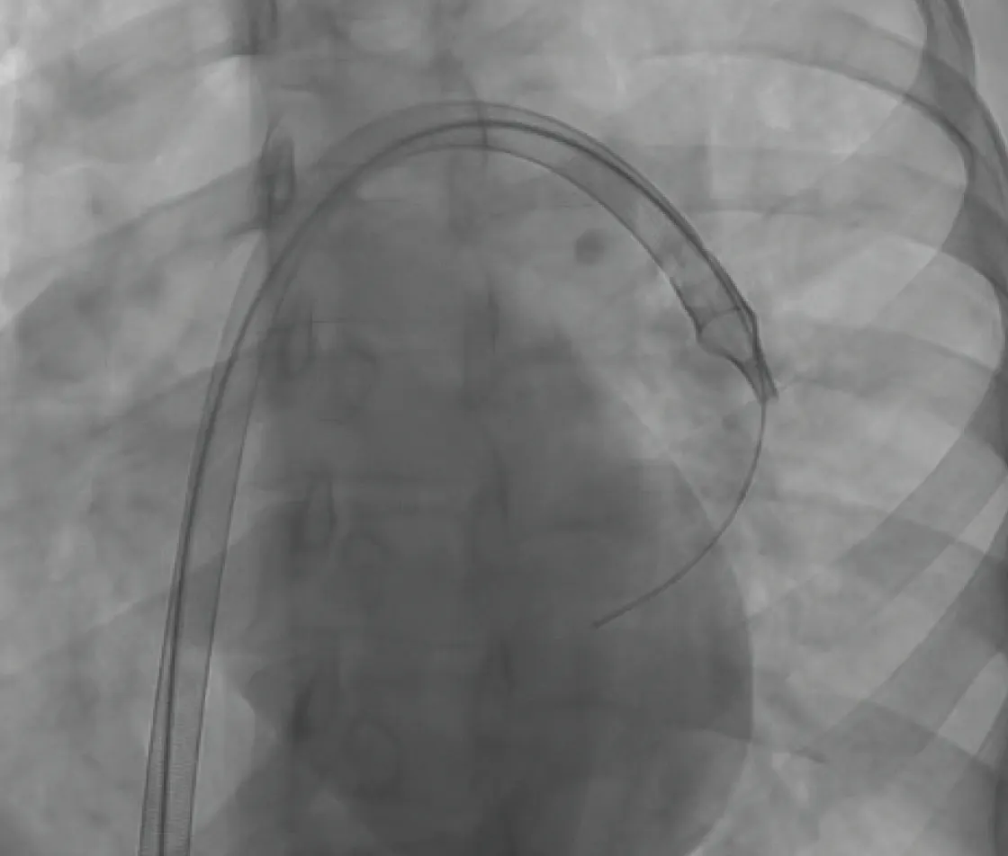
Figure 4. Directional AVENTUS Thrombectomy Catheter positioned to target the lower lobe of the left PA.
After treatment of the left PA, the AVENTUS Thrombectomy Catheter was pulled back into the main PA, and the guidewire was advanced into the distal right PA. With the aid of the integrated navigation catheter, the AVENTUS Thrombectomy Catheter was positioned in the mid right PA (Figure 5), where several aspirations quickly reduced the high clot burden seen in precase imaging.
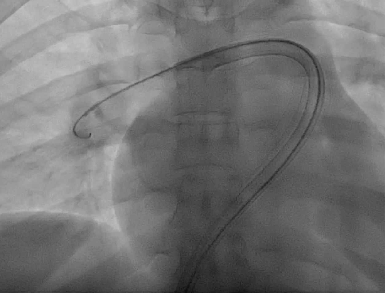
Figure 5. AVENTUS Thrombectomy Catheter positioned for initial aspirations in the right PA.
Then, 10 mL of contrast was injected into the fluid injection port of the AVENTUS Thrombectomy Catheter, and angiography demonstrated persistent thrombus within the TA branch (Figure 6).
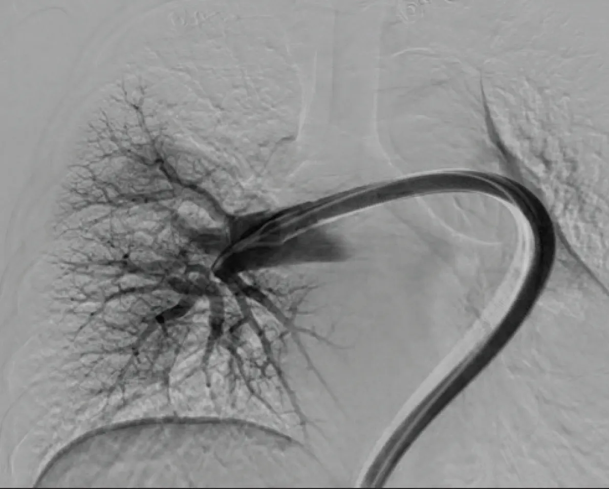
Figure 6. Angiography showing occluded TA of the right PA.
Rather than repositioning the guidewire or advancing the aspiration catheter into the branch itself, the guidewire was maintained in its existing distal right PA position. The AVENTUS Thrombectomy Catheter was retracted to the ostium of the TA bifurcation and rotated using its directional aspiration capability until the aspiration port was oriented directly toward the branch ostium (Figure 7). A single aspiration pass successfully removed the residual TA thrombus without requiring guidewire repositioning or branch vessel catheterization. A final angiography image (Figure 8) confirmed successful aspiration of clot in the TA and restored perfusion throughout the right PA.
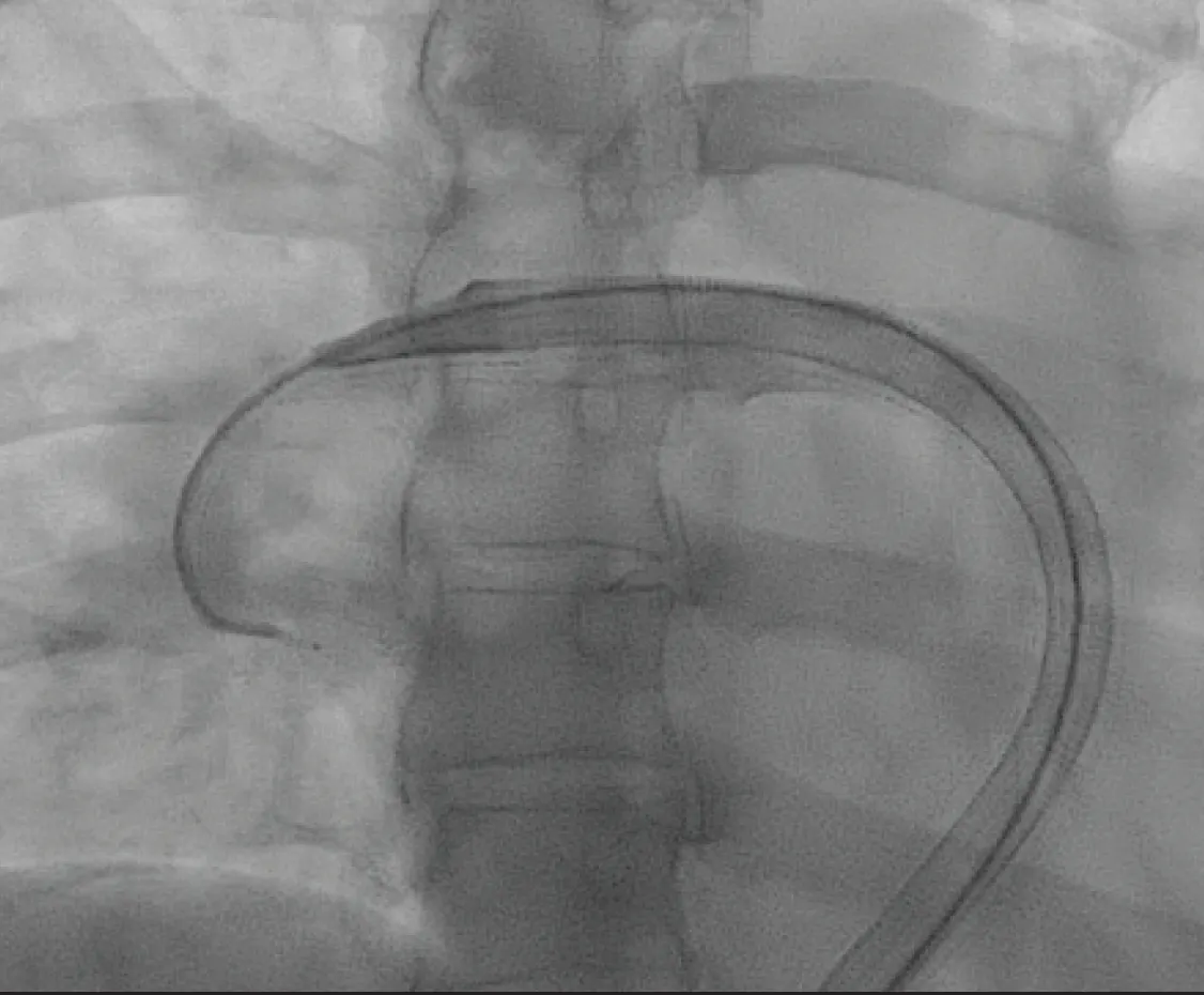
Figure 7. The AVENTUS Thrombectomy Catheter with its directional aspiration tip positioned to extract clot in the TA.
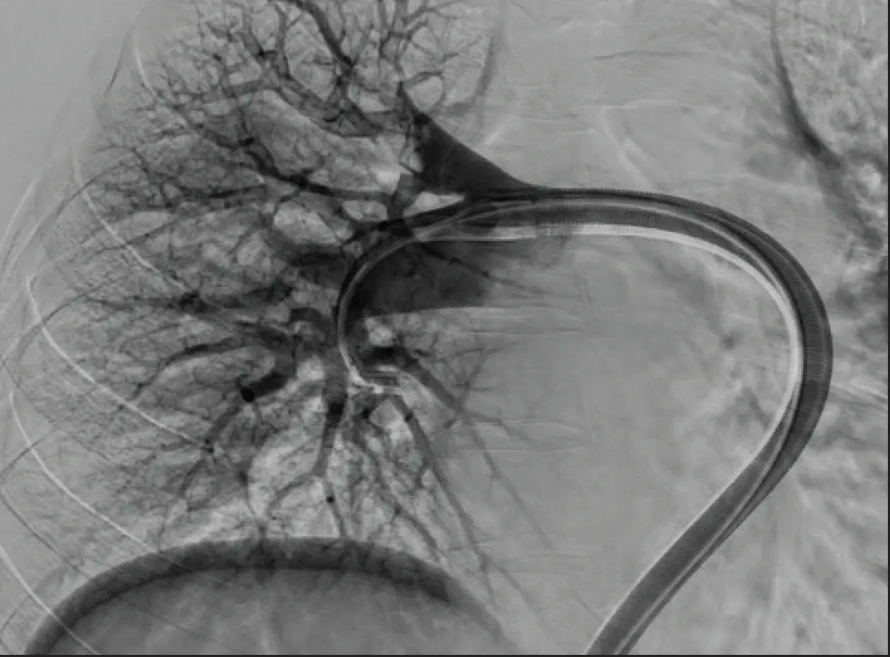
Figure 8. Final angiography showing perfusion throughout the right PA.
POSTINTERVENTION RESULTS
Overall, the total time from thrombectomy catheter insertion to complete resolution of clot was only 15 minutes. The patient experienced immediate clinical improvement after MT (Figure 9). Oxygen saturation improved from in the 80% range prior to intervention to 99% immediately after treatment. The patient left the procedure table breathing comfortably on room air and reported a marked improvement in symptoms before leaving the catheterization laboratory.
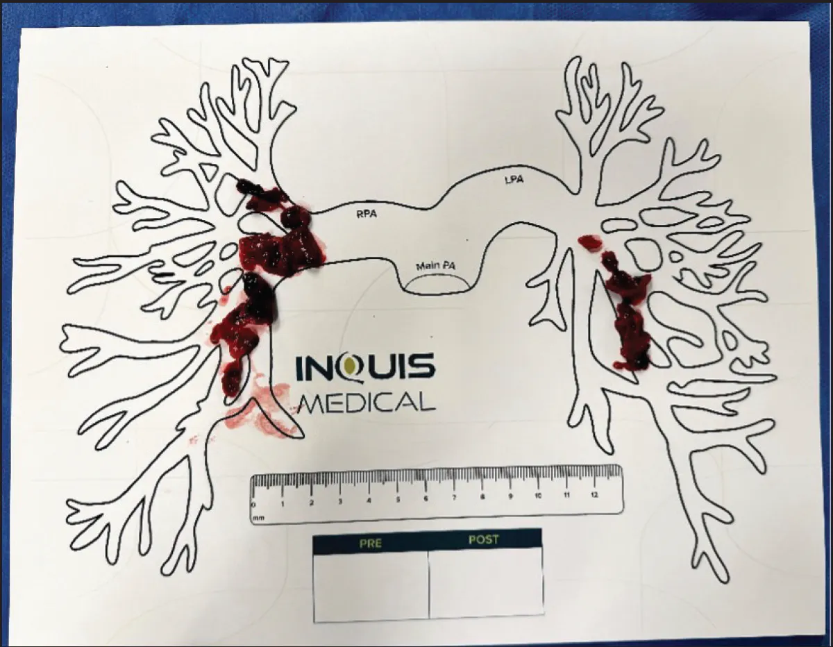
Figure 9. Thrombus extracted during the procedure.
DISCUSSION
This case highlights the value of directional aspiration during PE thrombectomy. Residual thrombus within branch vessel ostia can often require guidewire repositioning and additional catheter manipulation to achieve complete clot removal. In this patient, the directional aspiration capability of the AVENTUS Thrombectomy System enabled targeted aspiration of TA thrombus from the bifurcation, eliminating the need for guidewire exchange or advancement of the aspiration catheter into the branch.
The ability to precisely direct aspiration toward residual thrombus may simplify procedural workflow, reduce procedural steps, and facilitate treatment of branch vessel clot while maintaining stable guidewire position. Combined with immediate hemodynamic and symptomatic improvement, this case demonstrates how directional aspiration can enhance procedural efficiency while achieving effective clot removal in patients with intermediate-high–risk PE.

Indications for Use: The Aventus Thrombectomy System is indicated for:
- The non-surgical removal of emboli and thrombi from blood vessels.
- Injection, infusion, and/or aspiration of contrast media and other fluids into or from a blood vessel.
The Aventus Thrombectomy System is intended for use in the peripheral vasculature and for the treatment of pulmonary embolism.
Indications for Use: The Aventus Clot Management System is indicated for use with the Aventus Thrombectomy System for autologous blood transfusion.
Indications for Use: The Aventus Introducer Sheath is intended to be inserted in the vasculature to provide a conduit for the insertion of endovascular devices while minimizing blood loss associated with such insertions.
Advertisement
Advertisement