Advertisement
Advertisement
May 2026
Applicability of OCT in Below-the-Knee Percutaneous Transluminal Angioplasty
Rationale for use of optical coherence tomography during BTK intervention, procedural steps, limitations, and future considerations.
By Chrissy van Wely, MD; Rens J. Oosterveld, MD; Lieke C.D. Brugman, MSc; Ozan Yazar, MD, PhD; and Lee H. Bouwman, MD, PhD
Peripheral artery disease is a manifestation of atherosclerosis that is associated with substantial morbidity and mortality, particularly in advanced cases such as chronic limb-threatening ischemia (CLTI).1 Patients with CLTI are at an increased risk of major amputation as well as elevated cardiovascular morbidity and mortality rates.2 Below-the-knee (BTK) arterial disease, frequently the underlying pathology in patients with CLTI, is characterized by small-caliber vessels with heavily calcified lesions, resulting in technically challenging revascularization procedures.3,4
The current gold standard imaging modality to guide percutaneous transluminal angioplasty (PTA) is digital subtraction angiography (DSA); however, it has significant inter- and intraobserver variability.5 These limitations, combined with the low patency rates after BTK revascularization procedures, emphasize the need for more objective and reproducible imaging techniques.
Optical coherence tomography (OCT) is an intravascular imaging technique that provides high-resolution, cross-sectional visualization of the vessel wall, enabling detailed assessment of plaque morphology and accurate measurement of vessel dimensions. OCT catheters emit near-infrared light into the vessel, which is absorbed, backscattered, or reflected by tissue structures. The reflected signal is analyzed using interferometry to generate high-resolution images with an axial resolution of 10 to 20 µm, which is approximately 10 times higher compared to other intravascular imaging techniques such as intravascular ultrasound (IVUS).6
APPLICABILITY IN BTK INTERVENTIONS
The use of OCT in the peripheral arteries is currently outside instructions for use and remains limited to investigational settings. Despite the small evidence base, early studies show promising results with the use of OCT in BTK arteries. OCT has been shown to be safe and feasible to use in BTK arteries without any major complications.7 Importantly, OCT can detect (major) dissections and flow-limiting thrombus that would otherwise not be seen using only DSA (Figure 1). In a small proof-of-principle study of 10 patients, perioperative decision-making based on OCT would have changed the physician’s treatment strategy in 20% of patients due to thrombus or dissections. Additionally, there was a discrepancy between vessel diameter measured with OCT versus DSA.7 These findings are similar to the widely known evidence from percutaneous coronary artery intervention.8
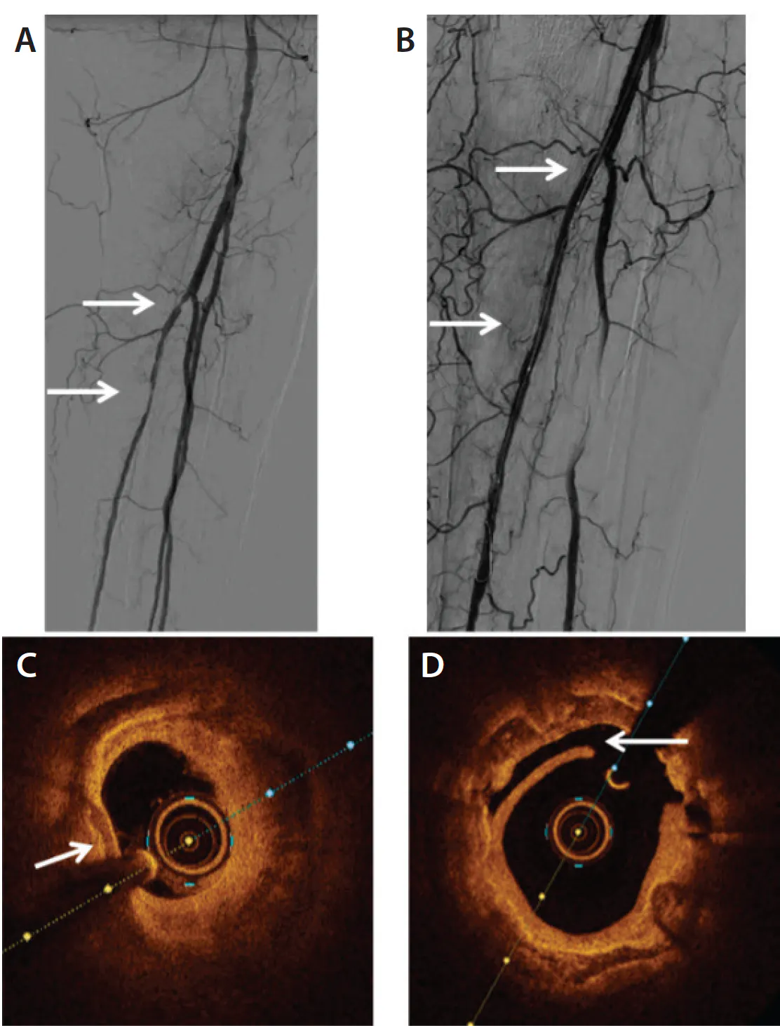
Figure 1. DSA and OCT images of a patient treated with PTA.Preintervention angiography with stenosis in the proximal posterior tibial artery (A). Postintervention angiography without residual stenosis and no visible thrombosis or dissection (B). Preintervention OCT image demonstrating heavily calcified plaque (arrow) with a severe loss of lumen (C). Postintervention OCT image demonstrating a widened lumen as compared to the preintervention image and dissection (arrow) (D). Reprinted from van Wely C, Oosterveld RJ, Bouwman LH, et al. Optical coherence tomography and fractional flow reserve in below-the-knee percutaneous transluminal angioplasty: a pilot study. CVIR Endovasc. 2025;8:87. doi: 10.1186/s42155-025-00580-9
The importance of accurate vessel measurement is emphasized with BTK stents and scaffolds.9 Because vessel diameter appears to be frequently underestimated based on DSA, intravascular imaging techniques such as OCT can be used to determine the correct diameter and ensure accurate scaffold sizing. A larger scaffold may then be chosen based on OCT images before scaffold placement. Figure 2 shows angiograms before and after scaffold placement. Scaffold placement was performed using OCT guidance and resulted in the decision to increase scaffold sizing versus the original plan based on DSA. OCT images obtained during the procedure are shown in Figure 3. Additionally, post–scaffold placement OCT images showed malapposition of the scaffold, which was not visible on DSA. Postdilatation was performed with a semicompliant balloon, and OCT subsequently showed good apposition to the vessel wall and adequate expansion. This demonstrates that angiography by itself fails to show malapposition of the scaffolds in BTK arteries, whereas OCT precisely shows where apposition may be suboptimal and additional dilatation is required to improve patency rates (Figure 2 and Figure 3).

Figure 2. Angiograms before (A) and after (B) scaffold placement and postdilation with the Esprit BTK system (Abbott).
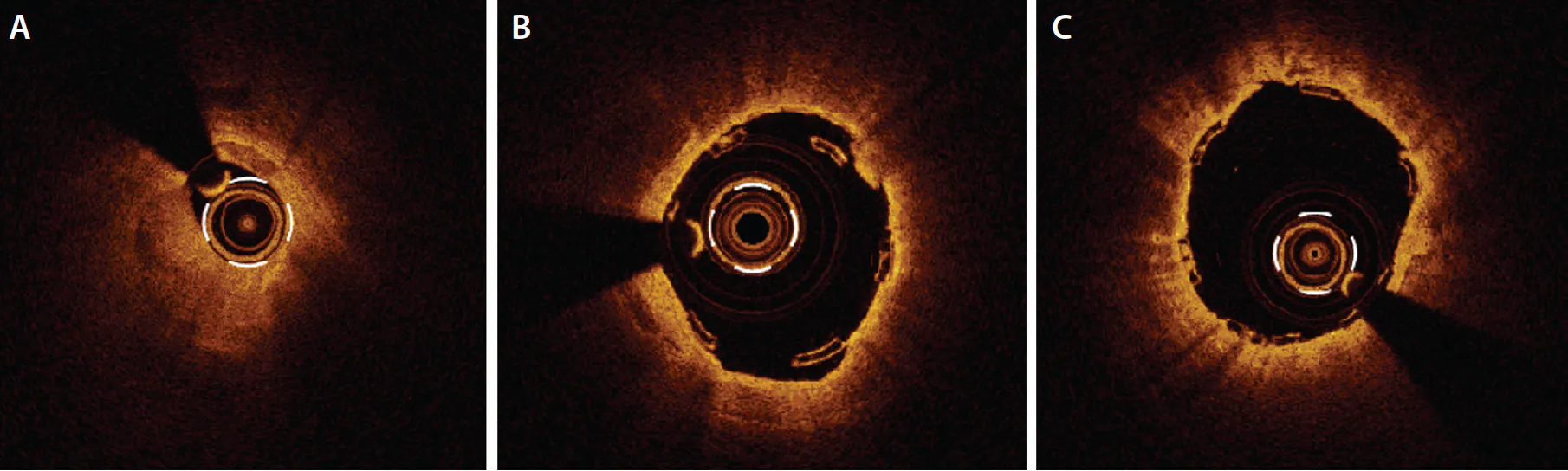
Figure 3. Cross-sectional OCT images of a patient treated with a scaffold in the anterior tibial artery. Preintervention OCT image demonstrating a near occlusion (A). Malapposition of the scaffold after placement (B). OCT image after postdilation demonstrating good apposition and expansion of the scaffold (C).
PROCEDURAL STEPS UTILIZING OCT FOR BTK INTERVENTIONS
The procedure may be performed under local or general anesthesia. Access is obtained via an ultrasound-guided antegrade puncture of the common femoral artery. Protocols for OCT catheters may vary based on the guidewire or sheath selected. In general, a 0.014-inch guidewire is introduced, and a bolus of intravenous heparin is given based on hospital protocol. A sheath of at least 6 F is introduced, and DSA is performed to visualize the femoropopliteal and crural arteries. A guiding catheter is not required to obtain OCT measurements but may be used at the physician’s discretion. If the physician opts for a guiding catheter, it should be at least 6 F. The OCT catheter has a hydrophilic coating to ensure smooth delivery of the catheter past the lesion of interest. Based on the DSA, the lesion of interest is chosen, and the OCT catheter is placed in the correct position, guided by its radiopaque markers. A bolus of 16 mL pure iodine contrast agent is used to flush blood from the target vessel, enabling clear OCT image acquisition during the automatic pullback. Images are processed either through an integrated angiography suite system or via a mobile OCT console. The software automatically measures the lumen and external elastic lamina but also allows the physician to manually edit these measurements if necessary. When integrated into the angio suite, angiography coregistration enables precise localization for balloon angioplasty or scaffold placement.
LIMITATIONS
The need for blood clearance using an iodine contrast agent might be used as an argument against using OCT to guide PTA, particularly in a patient population that often has preexisting renal impairment. Although OCT-guided revascularization procedures indeed require more contrast compared to revascularization procedures guided by other imaging modalities such as IVUS, a higher incidence of contrast-induced nephropathy has not been noted in OCT-guided interventions.10 Although alternative methods to facilitate blood clearance exist (eg, heparinized saline, Dextran, CO2), further research must be conducted to compare their efficacy and safety with the manufacturer-recommended use of pure iodine contrast agent.11
Furthermore, OCT catheters were designed for use in the coronary arteries and are therefore relatively short. This results in limited use of OCT in BTK arteries, as cross-over interventions are impossible due to the short catheter length.12 Future innovations and alterations in catheter design and length may facilitate cross-over interventions in the future.
FUTURE DIRECTIONS
There is a shift toward intravascular imaging–guided BTK revascularization procedures. OCT adds value in terms of sizing and planning BTK revascularization procedures, particularly when scaffolds are implanted. Further research is required to define its exact added value in standard BTK revascularization procedures and to compare its performance to other intravascular imaging techniques, including IVUS. Prospective studies evaluating long-term clinical outcomes of OCT- versus angiography-guided BTK interventions are warranted.
1. Conte MS, Bradbury AW, Kolh P, et al. Global vascular guidelines on the management of chronic limb-threatening ischemia. Eur J Vasc Endovasc Surg. 2019;58:S1-S109.e33. doi: 10.1016/j.ejvs.2019.05.006
2. Hata Y, Iida O, Asai M, et al. Risk stratification for 2-year mortality in patients with chronic limb-threatening ischemia undergoing endovascular therapy. J Atheroscler Thromb. 2021;28:477-482. doi: 10.5551/jat.57711
3. Kleiss SF, van Mierlo-van den Broek PAH, Vos CG, et al. Outcomes and patency of endovascular infrapopliteal reinterventions in patients with chronic limb-threatening ischemia. J Endovasc Ther. 2024;31:831-839. doi: 10.1177/15266028221147457
4. Snyder DJ, Zilinyi RS, Pruthi S, et al. Percutaneous transluminal angioplasty for infrapopliteal chronic limb-threatening ischemia: a systematic review and meta-analysis of primary patency and binary restenosis rates. J Endovasc Ther. 2025;32:1547-1561. doi: 10.1177/15266028231212133
5. van Wely C, Oosterveld RJ, Zonnebeld N, et al. Interobserver and intraobserver variability in crural angiography. J Endovasc Ther. Published online January 24, 2025. doi: 10.1177/15266028251315029
6. Jiang Y, Wang J, Sreeramachandra Murthy R, et al. Intracoronary optical coherence tomography: technological innovations and clinical implications in cardiology. Curr Treat Options Cardiovasc Med. 2025;27:44. doi: 10.1007/s11936-025-01103-4
7. van Wely C, Oosterveld RJ, Bouwman LH, et al. Optical coherence tomography and fractional flow reserve in below-the-knee percutaneous transluminal angioplasty: a pilot study. CVIR Endovasc. 2025;8:87. doi: 10.1186/s42155-025-00580-9
8. Koros R, Karanasos A, Papafaklis MI, et al. Latest evidence on intravascular imaging: a literature review. J Clin Med. 2025;14:4714. doi: 10.3390/jcm14134714
9. DeRubertis BG, Varcoe RL, Krishnan P, et al. Drug-eluting resorbable scaffold versus balloon angioplasty for below-the-knee peripheral artery disease: 2-year results from the LIFE-BTK trial. Circulation. 2025;152:1076-1086. doi: 10.1161/CIRCULATIONAHA.125.075080
10. Kang DY, Ahn JM, Yun SC, et al. Optical coherence tomography-guided or intravascular ultrasound-guided percutaneous coronary intervention: the OCTIVUS randomized clinical trial. Circulation. 2023;148:1195-1206. doi: 10.1161/CIRCULATIONAHA.123.066429
11. Kendrick DE, Allemang MT, Gosling AF, et al. Dextran or saline can replace contrast for intravascular optical coherence tomography in lower extremity arteries. J Endovasc Ther. 2016;23:723-30. doi: 10.1177/1526602816657392
12. Galo J, Al-Qaraghuli A, Wallace R, et al. Intravascular imaging in peripheral arterial disease: a contemporary literature review. Eur Heart J Open. 2026;6:oeag016. doi: 10.1093/ehjopen/oeag016
Advertisement
Advertisement