Advertisement
Advertisement
May 2026
Sponsored by Shockwave Medical
Shockwave Javelin in Complex Limb Salvage
Early clinical experience and evolving role in BTK and pedal interventions.
Chronic limb-threatening ischemia (CLTI) remains one of the most challenging arenas in endovascular therapy. These patients often present with multilevel disease, prior interventions, and dense medial calcification that limits device deliverability. In this population, the ability to modify calcium safely and effectively is not simply a technical preference—it is the determinant of whether therapy can be delivered at all.
Over the past year, the introduction of the Shockwave Javelin Peripheral IVL catheter (Shockwave Medical–Johnson & Johnson Medtech), the first-of-its-kind Forward intravascular lithotripsy (IVL) Platform, has expanded my ability to treat heavily calcified below-the-knee (BTK) and below-the-ankle (BTA) lesions. My early experience with the catheter’s design aligns with the clinical reality of limb salvage: When calcium is the barrier, energy delivery beyond the catheter tip can be the difference between procedural success and limb loss.
CALCIUM AS THE PROCEDURAL GATEKEEPER
In BTK and pedal interventions, calcium is not merely a lesion characteristic—it is the variable that determines whether therapy can be delivered. Dense, circumferential, or nodular calcification in the tibials and pedal arch can prevent wire traversal, microcatheter support, balloon tracking, adequate vessel preparation, and delivery of definitive therapy. Traditional IVL balloons have transformed vessel preparation in larger vessels, but the extensive calcification encountered in the distal tibials and foot limit their use. In these segments, the challenge is not balloon expansion—it’s access. This is where Shockwave Javelin has a unique role.
NOVEL FORWARD IVL PLATFORM
Shockwave Javelin delivers ultrasonic acoustic pressure waves spherically from the emitter (Figure 1), modifying calcium ahead of the catheter and creating a path for device delivery in tight, heavily calcified segments where no balloon could be advanced.* The acoustic output is consistent with the Shockwave Balloon IVL platform, and with its favorable safety profile,1-3 it provides operators peace of mind when integrating Shockwave Javelin into existing workflows. In practice, this means that lesions previously considered “uncrossable” or “untreatable” now have a viable path to therapy.
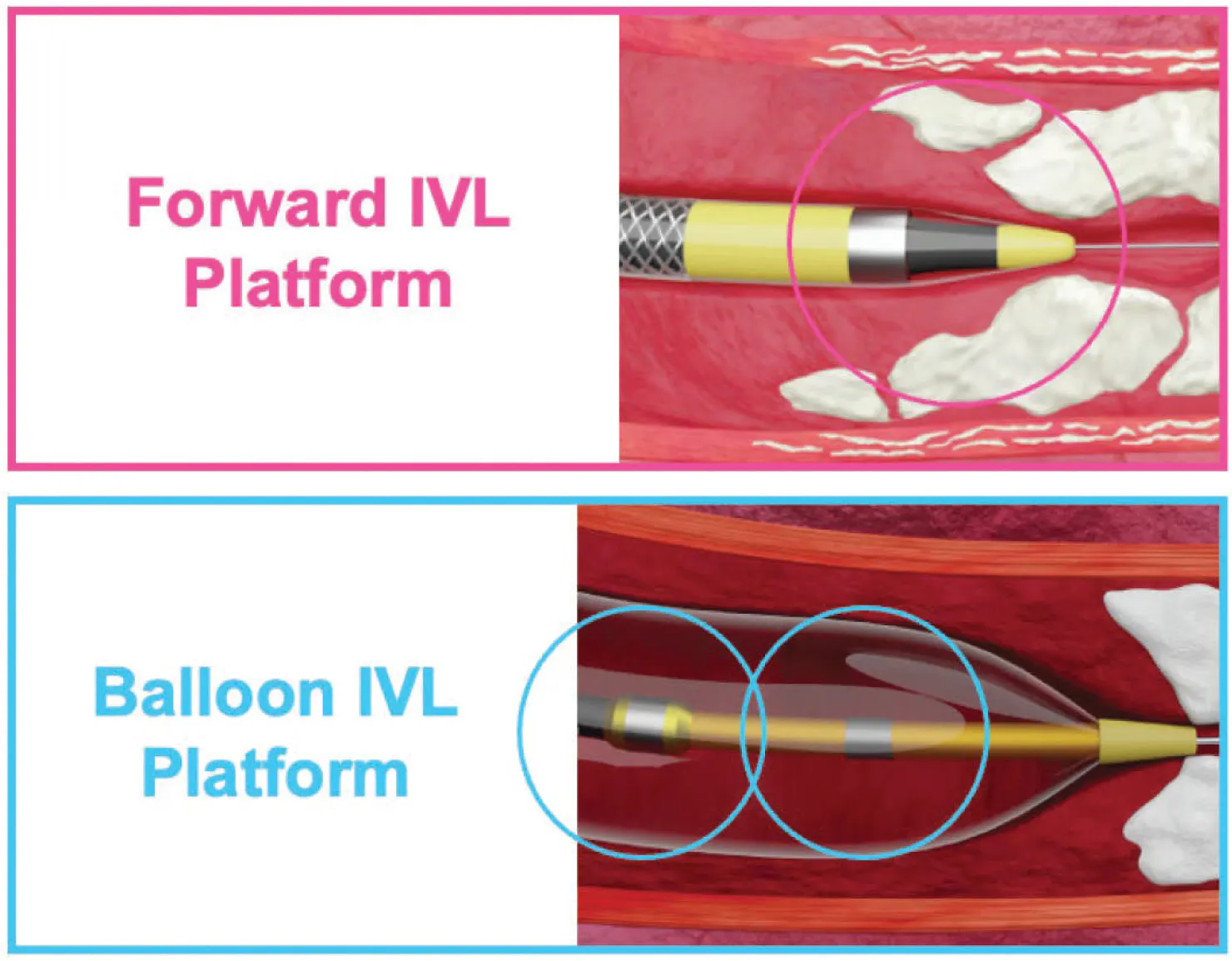
Figure 1. The sonic output of the Shockwave Forward IVL Platform compared to the Shockwave Balloon IVL Platform.
EARLY EXPERIENCE: FIRST 10 CONSECUTIVE CASES
My initial experience includes 10 consecutive BTK cases, three of which involved BTA disease. These were not selected cases; rather, they represent the full spectrum of CLTI complexity, including long-segment tibial occlusions, dense circumferential calcification, prior failed interventions, and pedal arch disease (Table 1).
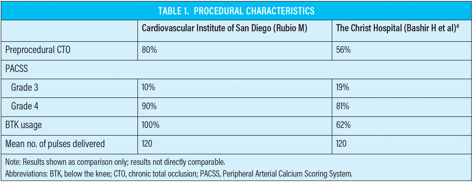
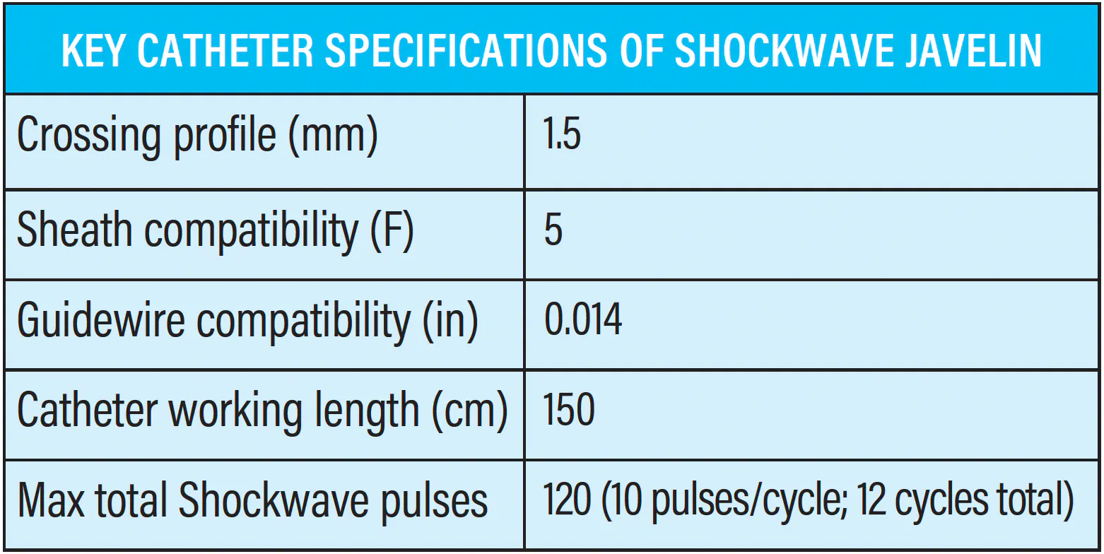
Across all 10 cases, the device demonstrated a favorable safety profile. I observed no perforations, no flow-limiting dissections, no distal embolization, and no device-related complications. Most cases required the full 120 pulses, which is consistent with the severity of calcification in this population. In two cases, the catheter reached its pulse limit before traversing the entire lesion, yet the calcium modification achieved was sufficient to allow balloon tracking and enable delivery of definitive therapy. This has been a consistent theme: Shockwave Javelin does not need to cross the entire lesion to change the procedural trajectory.
The three BTA cases highlight where Shockwave Javelin’s distal energy delivery has been most impactful in my practice. In two of these cases, the device enabled therapy in segments where no balloon or microcatheter could initially track. One case remained uncrossable, but this was a patient with end-stage vascular disease in whom no device—microcatheter or atherectomy platform—could cross. This reflects the biological limits of terminal disease rather than a device-specific limitation.
CASE SPOTLIGHT: RECANALIZATION OF THE DORSALIS PEDIS/FIRST DORSAL METATARSAL ARTERY
Case Presentation
A patient presented with a nonhealing right first-toe wound. Eight months earlier, they had undergone anterior tibial angioplasty, but the wound had failed to progress. On reevaluation, angiography demonstrated a chronic total occlusion of the dorsalis pedis artery (DPA) extending into the first dorsal metatarsal artery (DMA) (Figure 1).
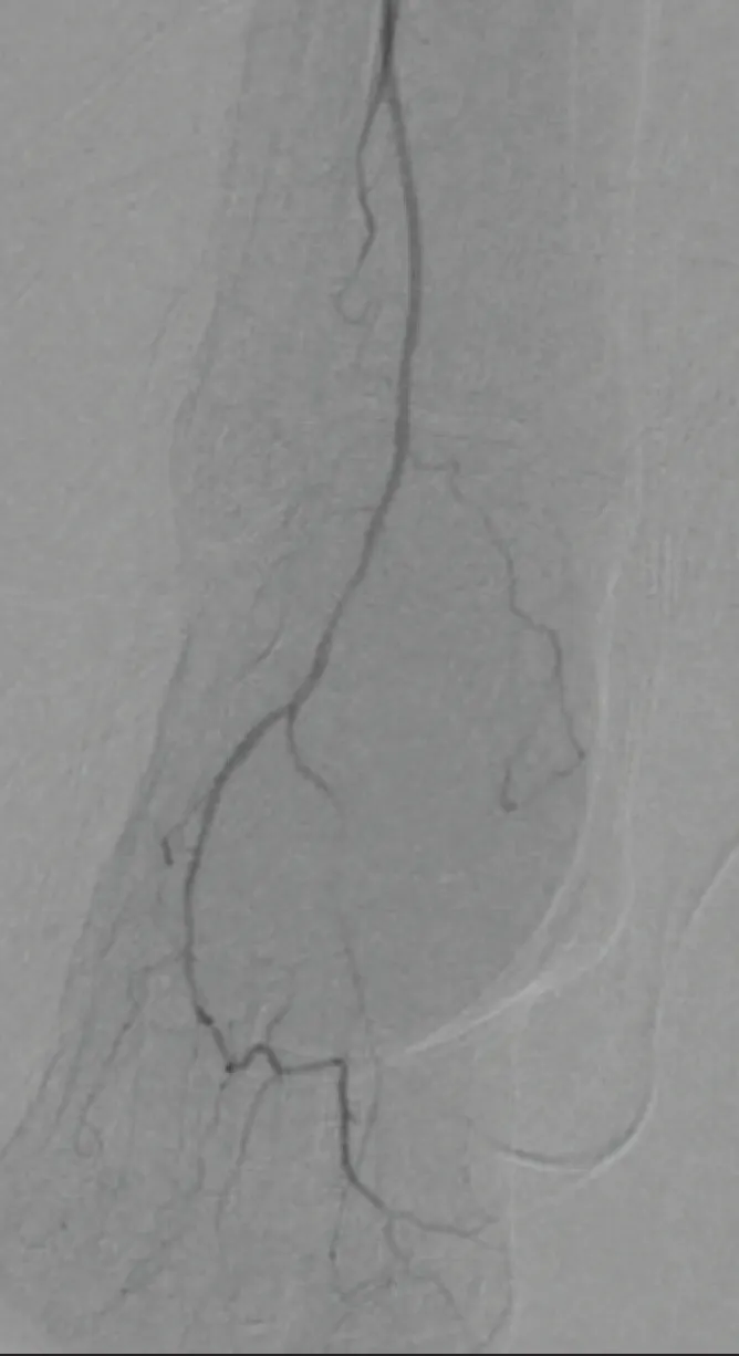
Figure 1. Occlusion of the DPA extending into the first DMA.
Procedural Overview
Vascular access was obtained via contralateral left common femoral artery, using a 6-F, 90-cm sheath, which provided adequate support. The occlusion was crossed with a 0.014-inch guidewire. An antegrade approach was not pursued, as the lesion was heavily calcified and unlikely to be more easily traversed. Rather than relying on increased forward force by antegrade approach, which risks subintimal passage or vessel injury, calcium modification was prioritized to enable treatment and subsequent device delivery.
Crossing the lesion with a wire was possible, but delivering therapy was not; the calcification in this distal segment was too dense. Shockwave Javelin was delivered at the level of maximal resistance, corresponding to the segment where the intravascular ultrasound (IVUS) catheter demonstrated mechanical “wedging” (Figure 2). IVUS revealed a heavily calcified, napkin-ring lesion at the exact point where the catheter could not advance further. The majority of the 120 pulses were delivered in this focal segment to optimize plaque modification (Figure 3). The remaining pulses were then applied more distally, within the proximal third of the lesion. This created enough compliance to allow delivery of a low-profile balloon (Figure 4), enabling successful recanalization of the DPA/first DMA (Figure 5). Perfusion improved immediately, and the wound—stagnant for 8 months—is now nearly healed.
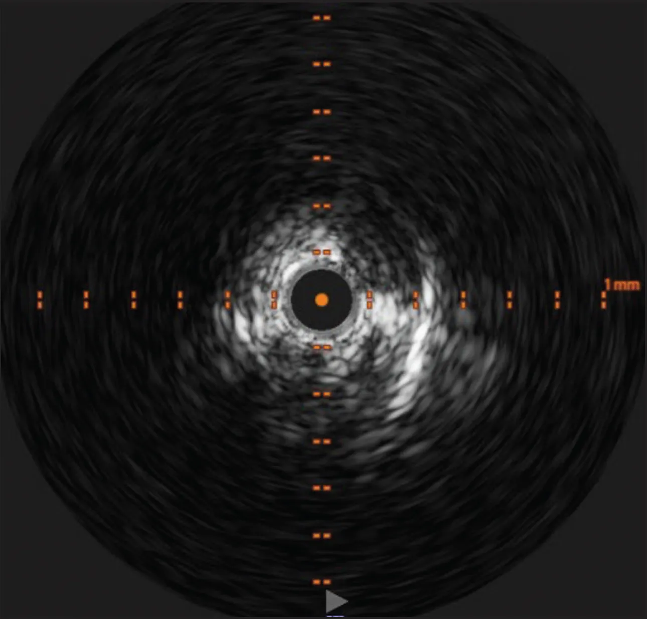
Figure 2. IVUS showing a heavily calcified, napkin-ring lesion at the point where the catheter was wedged.
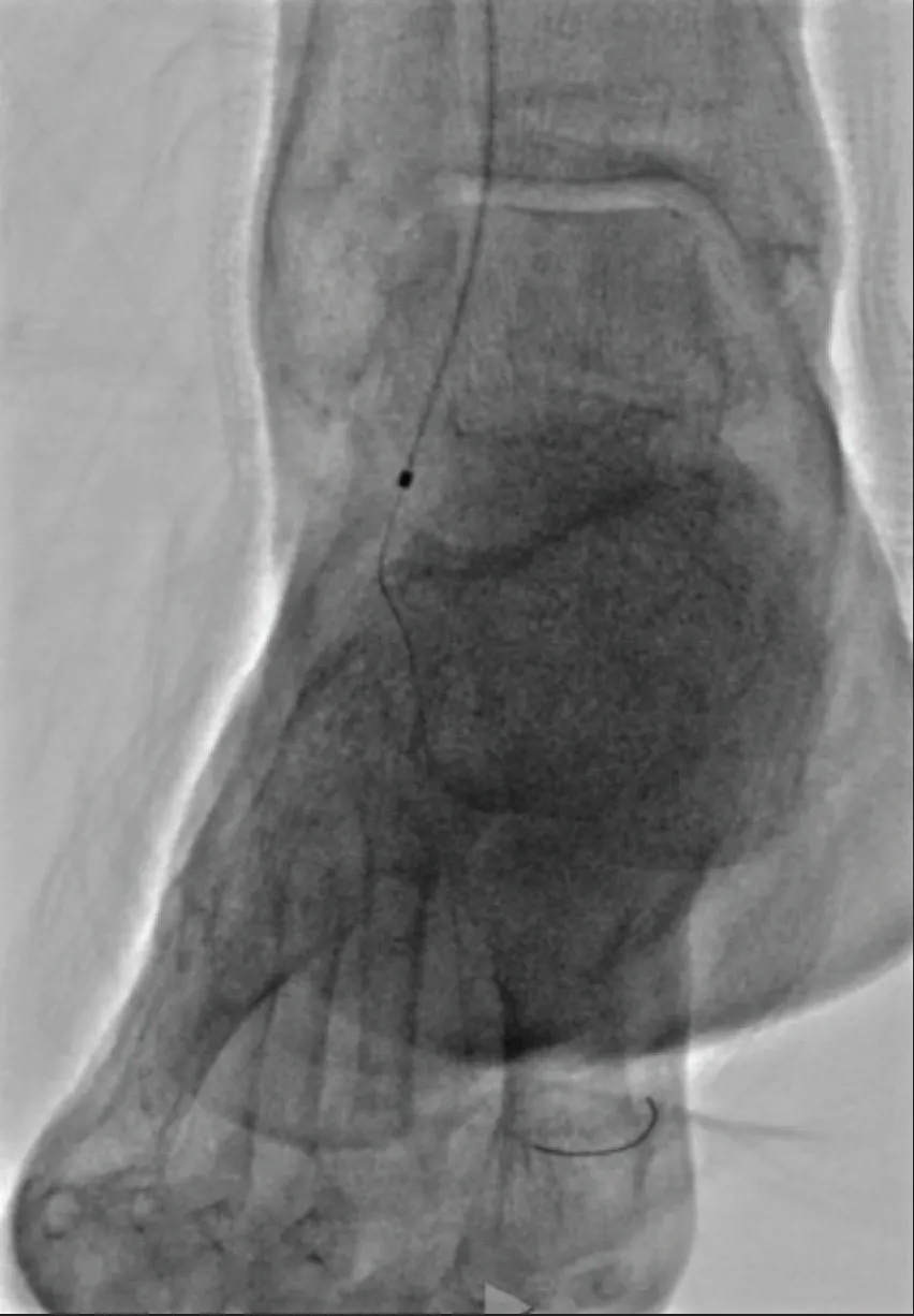
Figure 3. Shockwave Javelin catheter entering the occlusion.
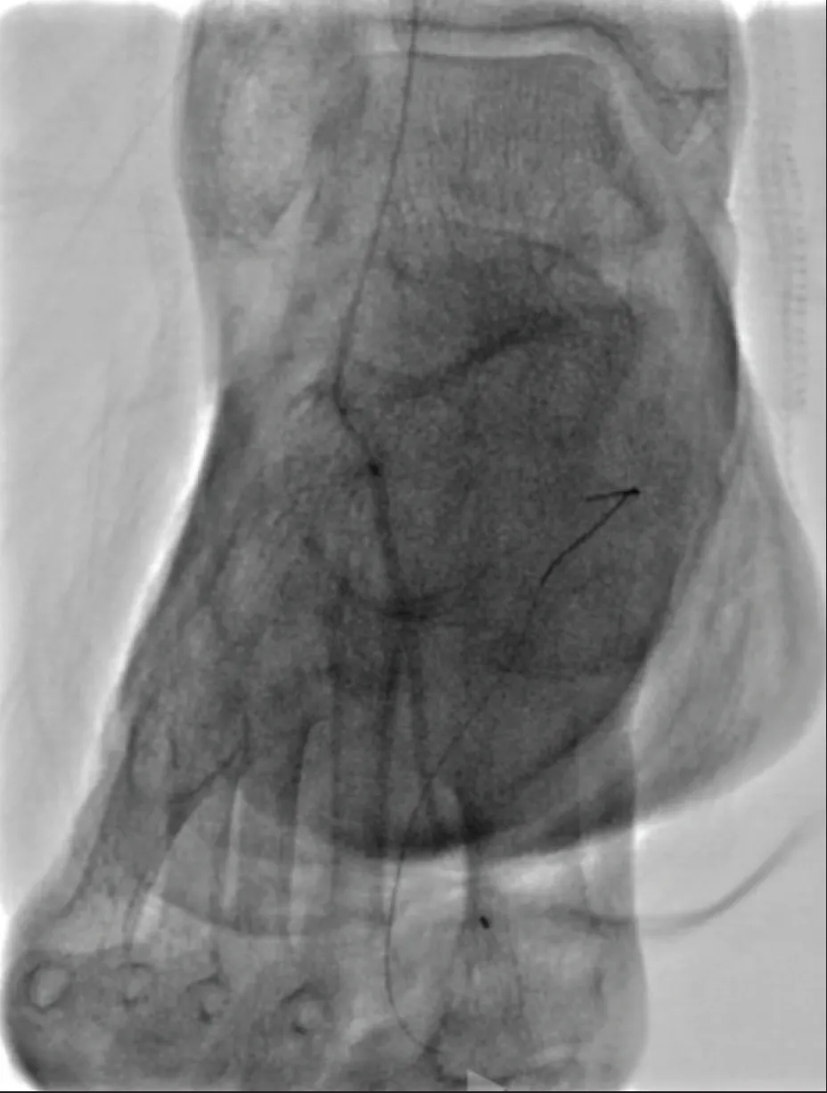
Figure 4. Delivery of low-profile balloon after calcium modification.
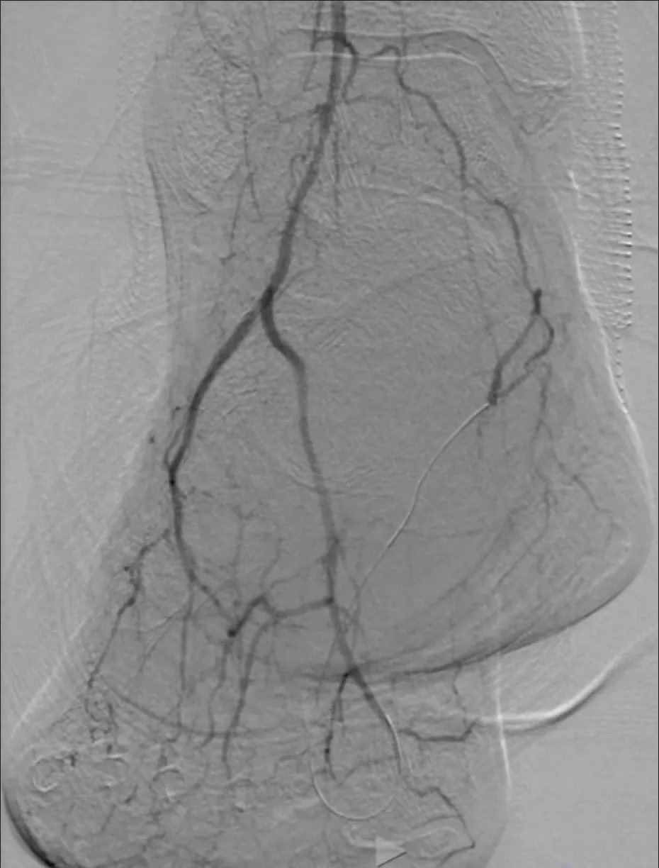
Figure 5. Successful recanalization of the DPA/first DMA.
This case illustrates how the novel Shockwave Forward IVL Platform can change the trajectory of a limb salvage procedure by enabling therapy in distal, heavily calcified anatomy where no other device can advance.
Clinical Decision-Making in Limb Salvage
Limb salvage requires a willingness to use every reasonable tool that may improve perfusion and preserve function. These patients often present with limited options, and procedural success frequently hinges on whether therapy can be delivered through heavily calcified segments. Shockwave Javelin is a new tool I can now reach for when calcium is the final barrier between the patient and a meaningful chance at limb preservation.
COMPARISON TO PUBLISHED EXPERIENCE
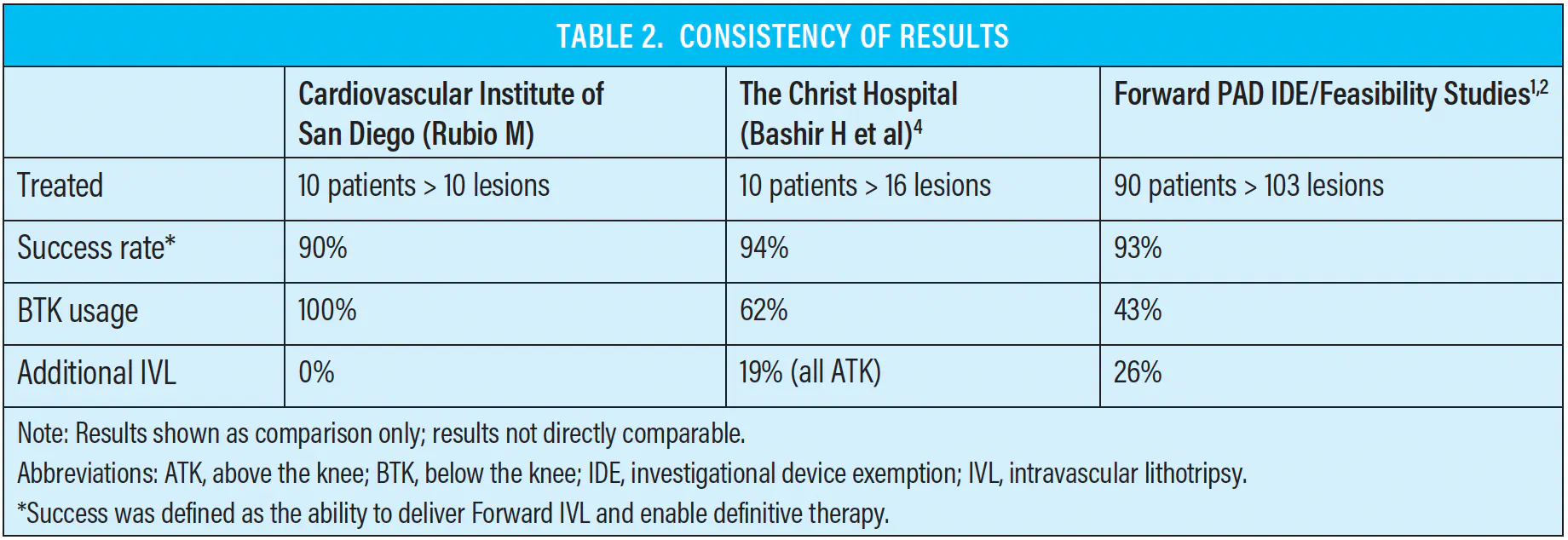
The initial 10-case institutional experience from The Christ Hospital in Cincinnati, Ohio, published by Bashir et al in JSCAI reported high rates of successful calcium modification, improved device delivery, procedural success in heavily calcified BTK lesions, and favorable safety outcomes.4 My early experience mirrors these findings (Table 2). Both series highlight that Shockwave Javelin’s value lies not in luminal gain, but in enabling therapy—transforming an uncrossable lesion into a treatable one.
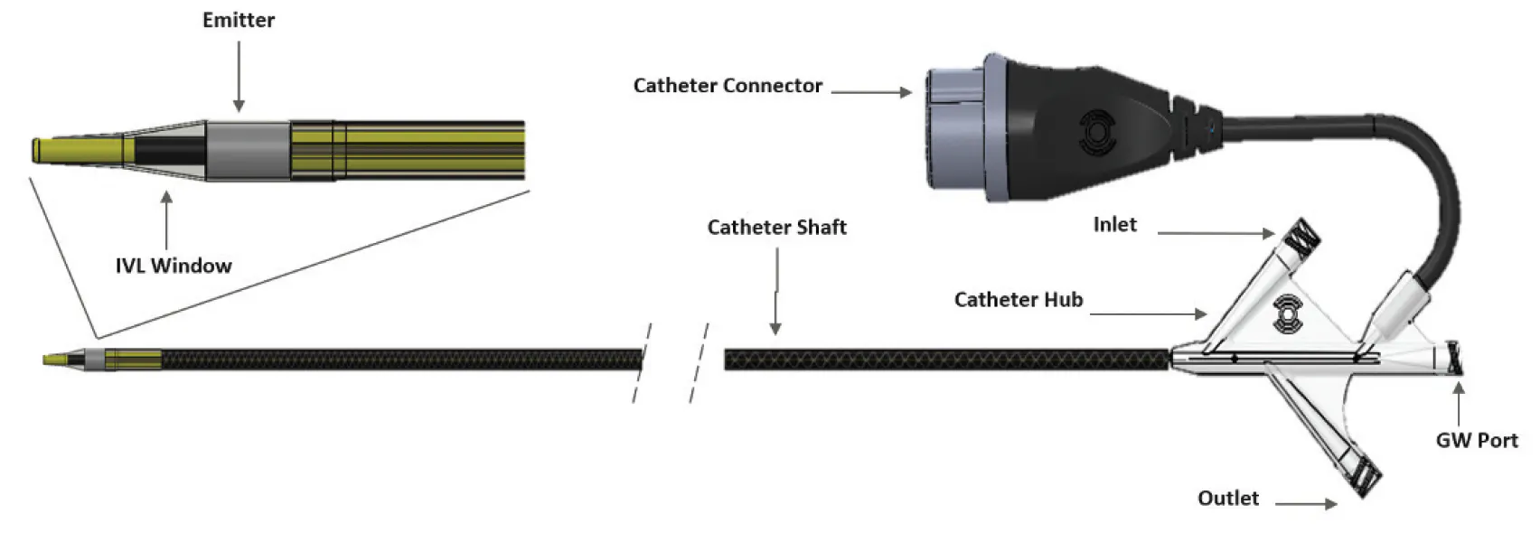
The components of the Shockwave Javelin Catheter.
CLINICAL PEARLS OF SHOCKWAVE JAVELIN
- Energy delivered beyond the tip is the differentiator
- Utilize all 120 pulses
- Partial traversal can still succeed
- BTK and BTA lesions benefit most
- Safety profile is favorable
CONCLUSION
Shockwave Javelin has expanded my ability to treat heavily calcified BTK and pedal lesions by modifying calcium beyond the tip, enabling device delivery where it was previously impossible, maintaining a strong safety profile, and aligning with the real-world needs of limb salvage. My early experience reinforces the device’s role in modern infrapopliteal intervention. Although no tool is perfect, Shockwave Javelin has repeatedly allowed me to deliver therapy in cases where calcium was the final barrier between a patient and limb preservation. In limb salvage, that difference matters.
*Internal testing shows a spherical sonic output from distal emitter consistent with previous Shockwave balloon-based IVL emitters, with sonic output extending beyond the tip of the catheter.
1. Corl JD, Clair D, Mwipatayi P, et al. FORWARD PAD IDE/feasibility studies: primary endpoint analysis of a novel non-balloon-based peripheral IVL catheter. JACC Cardiovasc Interv. 2025;18:398-399. doi: 10.1016/j.jcin.2024.10.035
2. Corl J. First clinical use of an innovative forward-shifted peripheral intravascular lithotripsy system: late-breaking primary outcomes of the mini S/FORWARD PAD IDE study. Presented at: Vascular Interventional Advances (VIVA) 2024; November 4, 2024; Las Vegas, Nevada.
3. Armstrong E. One-year outcomes from the Disrupt PAD BTK II study: treatment of patients with calcified below-the-knee lesions with a peripheral intravascular lithotripsy system. Presented at: Vascular Interventional Advances (VIVA) 2025; November 3, 2025; Las Vegas, Nevada.
4. Bashir H, Wong A, Muuse J, et al. Calcium modification during peripheral intervention with a novel intravascular lithotripsy system: an institutional experience. J Soc Cardiovasc Angiogr Interv. 2025;4:103708. doi: 10.1016/j.jscai.2025.103708

Shockwave Javelin Peripheral IVL Catheter Safety Information
In the United States: Rx only.
Indications for Use—The Shockwave Medical IVL System with the Javelin Peripheral IVL Catheter is intended for lithotripsy- enabled modification and crossing of calcified lesions in the peripheral vasculature, including the iliac, femoral, ilio-femoral, popliteal, and infra-popliteal arteries, prior to final treatment.
Not for use in coronary, carotid, or cerebral vasculature. Additionally, not for use in pulmonary vasculature in the U.S. and New Zealand.
Contraindications—Do not use if unable to pass 0.014″ (0.36 mm) guidewire across the treatment site-Not intended for treatment of in-stent restenosis or in coronary, carotid, cerebral or pulmonary arteries.
Warnings—Only to be used by physicians who are familiar with interventional vascular procedures—Physicians must be trained prior to use of the device— Use the generator in accordance with recommended settings as stated in the Operator’s Manual.
Precautions—Avoid applying acoustic pressure pulses while IVL window is not filled with sterile saline—Appropriate anticoagulant therapy should be administered by the physician— Decision regarding use of distal protection should be made based on physician assessment of treatment lesion morphology.
Adverse effects—Possible adverse effects consistent with standard angioplasty include-Access site complications-Allergy to contrast or blood thinner-Arterial bypass surgery—Bleeding complications—Death—Fracture of guidewire or device—Hypertension/Hypotension—Infection/sepsis—Placement of a stent—renal failure— Shock/pulmonary edema—target vessel stenosis or occlusion—Vascular complications. Risks unique to the device and its use—Allergy to catheter material(s)—Device malfunction or failure.
Prior to use, please reference the Instructions for Use for more information on indications, contraindications, warnings, precautions and adverse events. www.shockwavemedical.com/IFU
© Shockwave Medical, Inc. All Rights Reserved. SPL 80269 Rev. A.
Dr. Rubio is a paid consultant to Shockwave Medical.
Advertisement
Advertisement