Advertisement
Advertisement
June 26, 2023
Cook Medical’s Nester Fibered Coils Evaluated for Venous Use in Animal Study
June 26, 2023—Cook Medical announced the recent publication of a study that evaluated the embolization performance of the company’s fibered Nester coils versus nonfibered coils in a venous setting in an ovine study. The article, sponsored by Cook, was published by White et al in Journal of Vascular and Interventional Radiology (JVIR).
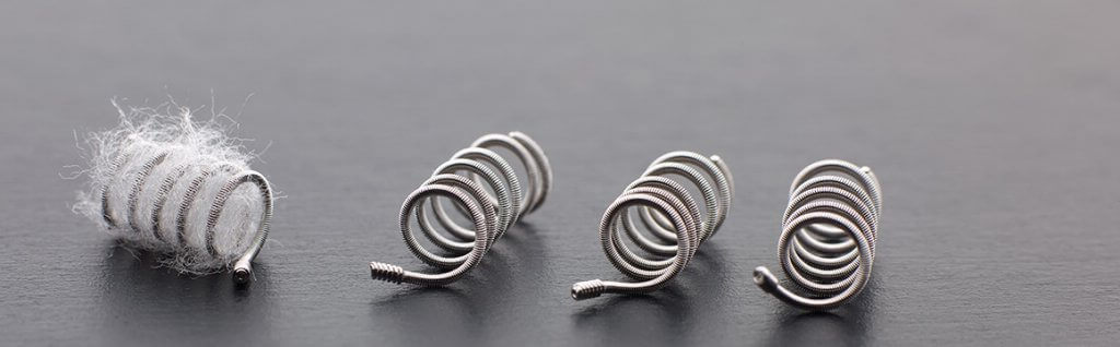
Researchers deployed fibered Nester coils and nonfibered coils in 24 veins in six sheep.
The company noted that investigators in the study found that fibered coils demonstrated several advantages over the nonfibered coils. Stasis was achieved in 5.3 minutes when the fibered coils were used versus 9 minutes for veins treated with nonfibered coils. Additionally, a lower average radiation dose was observed when using the Nester fibered coils (25.3 vs 34.9 mGy).
This research builds upon previous data that had showed similar advantages of fibered coils when compared with nonfibered coils in an arterial setting. The previous study, also sponsored by Cook, was published in 2019 by Trerotola et al in JVIR.
In their announcement, Cook noted that in both studies, fewer fibered coils were necessary to achieve acute occlusion versus bare metal coils. On average in the recent venous study, 5 (70 cm) fibered coils were needed to achieve occlusion versus 8.75 (122.5 cm) coils of bare metal coils. Comparatively, on average in the arterial study 1.3 (9.1 cm) fibered coils were needed to achieve occlusion versus 3.2 (22.4 cm) of nonfibered coils.
 Cook also highlighted that as part of the new venous fiber study, a novel approach to generating histologic images was used. Developed by Cook Research Incorporated and Alizée Pathology, to create histologic images cutting 5 µm thin sections of tissue with metallic implants. According to the company, thin sectioning results in clearer and more detailed histologic images, allowing a pathologist to read and interpret tissue changes. An example of this image output can be seen to the right.
Advertisement
Advertisement
