Advertisement
Advertisement
November 2024 Supplement
Sponsored by Surmodics, Inc.
Building a Radial-First Peripheral Intervention Program at Baylor Scott & White The Heart Hospital—Plano
A conversation with Sameh Sayfo, MD, MBA, FSCAI, FACC; Eleanor Huff, MSN, RN, FACC; and Morgan Weems, RCIS.
In 2023, Baylor Scott & White The Heart Hospital—Plano, in Plano, Texas, became one of the first four hospitals nationwide to be verified as part of the Vascular Verification Program (Vascular-VP), a quality program launched by the American College of Surgeonswith the Society for Vascular Surgery.1 The hospital has a long-standing commitment to radial access as a quality metric for percutaneous coronary interventions (PCIs); today, 67% of its PCI cases are performed from the wrist. In the recent years, the team at Heart Hospital has adopted radial access for peripheral procedures, driven by the improved safety and patient satisfaction of the radial approach amply demonstrated in the medical literature.2-4
In close partnership with hospital administration and staff, interventional cardiologist Sameh Sayfo, MD, has led the facility’s radial-to-peripheral initiative. Dr. Sayfo, who is Endovascular Fellowship Program Director at the hospital, now performs 63% of peripheral procedures via radial access, with an annual infrainguinal case volume of approximately 150. We spoke with Dr. Sayfo; registered cardiovascular invasive specialist, Morgan Weems; and Vice President of Cardiovascular Service Line Operations, Ellie Huff, about radial-to-peripheral adoption at Baylor Scott & White The Heart Hospital—Plano and its impact on patient care.
KEY TAKEAWAYS*
- New tools allow for more routine use of radial access for peripheral interventions
- Partnering with administration can facilitate radial-peripheral adoption
- Evidence challenges traditional objections to the radial-peripheral approach
- The Sublime™ Radial Access Platform includes Sublime™ 5 Fr radial-length guide sheaths, ≤ 200 cm Sublime™ torqueable microcatheters, and Sublime™ PTA catheters suitable for BTK treatment
* Based on current practice and clinical experience of Sameh Sayfo, MD.
DRIVERS OF RADIAL-PERIPHERAL ADOPTION
As an interventional cardiologist, what led you to focus on peripheral procedures?
Dr. Sayfo: I was trained at Brown University under one the best endovascular interventionalists there is, Dr. Peter Soukas. Since then, I’ve had a strong interest in endovascular peripheral therapy. I feel as though patients with peripheral artery disease (PAD) in the United States don’t get the high quality of care they deserve, especially compared with the care given to coronary artery disease patients. A lot of people with PAD aren’t being diagnosed or offered therapy.5 Because of that, many are living in pain or lose their limbs. The data for amputation in the United States are very concerning. Many people who end up getting an amputation have never had a prior arterial examination and given the increased mortality rate in patients with amputation,6 PAD management has become a passion of mine.

Baylor Scott & White The Heart Hospital—Plano. Courtesy of Baylor Scott & White The Heart Hospital–Plano.
Regarding radial-to-peripheral procedures, our interest really picked up in 2020, during the COVID era. We didn’t have enough beds to accommodate the huge influx of patients. Patients didn’t want to stay in hospitals because they didn’t consider them safe. Radial access helped us get them home safely sooner, with fewer overnight stays.
How would you describe the patient population you serve?
Dr. Sayfo: Since I began my practice, I’ve seen increasing numbers of older patients coming in with more complicated disease. Many have had prior procedures such as aortobifemoral bypass or endovascular aneurysm repair (EVAR). The more complex the patient, the more likely they have had multiple groin access procedures and groin complications, and they’re often very hesitant to have another groin access procedure. They might be afraid to even see a doctor. With obesity in the United States reaching 40% recently,7 we are seeing more patients with body mass index (BMI) > 35 kg/m2, and we know that higher BMI equates to higher risk of groin access complications.8
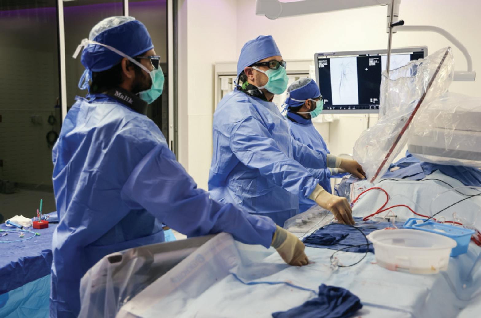
A radial-to-peripheral procedure underway at Baylor Scott & White The Heart Hospital—Plano. From left: M. Kasim Malik, DO, general cardiology fellow; Sameh Sayfo, MD, interventional cardiologist; and Usman Sarwar, MD, endovascular fellow. Courtesy of Baylor Scott & White The Heart Hospital–Plano.
Why are some patients so put off by groin access?
Ms. Weems: Groin access can involve a fair amount of discomfort and pain for patients.9 We use a local anesthetic during groin procedures, but local anesthesia is not always sufficient to manage the pain. When we deploy a vascular closure device, we have to give patients a warning and give them more pain medication, because it often hurts. When we need to apply manual compression, that takes 20 minutes even if things go well. If there’s any track ooze or if we have to give lidocaine-epinephrine to the groin, the patient is lying on their back a lot longer, dealing with the pressure we’re holding on their groin and the associated pain, which is worse if they have existing back pain.
I’ve seen patients with bruising all over their groins from previous cases. Some people have said their groin was very sore for a week after their femoral access procedure. Older patients who are on blood thinners or have fragile skin may have some bruising on their wrist from radial access, but it’s nothing compared with groin bruising.
How does recovery differ for femoral versus radial access?
Ms. Weems: With groin access, patients have to lie with their heads flat on the pillow for 2 to 3 hours with both legs straight. They want to be able to lift their heads, but we have to tell them that doing so applies pressure to the groin and they shouldn’t do it. If we go radial access, we just wake up the patient and their bed is ready right next to them. They just scoot over, and they can sit up right away. That makes a big difference to them.
When did you begin doing radial-to-peripheral procedures at the Heart Hospital?
Dr. Sayfo: Before 2019, we didn’t use radial access because we lacked radial-to-peripheral sheaths. For alternative access we’d sometimes use brachial or axillary, but I stayed away from them unless they were absolutely necessary because of complication risk.10,11 For alternative access we mainly used pedal access, which has advantages and disadvantages.12 In my opinion, the main disadvantage is if there is a superficial femoral artery (SFA) occlusion, the only angiogram being performed is distal to the occlusion, which prevents you from recognizing the complexity and length of the occlusion. So, you’re flying blind, trying to fix something based on a CT scan or ultrasound.
When Terumo Interventional Systems came out with radial-to-peripheral sheaths, I started tackling simple cases or cases that absolutely required radial access. Back then, I was doing about 10% of peripheral cases radially. But over the years, my volume of radial-to-peripheral cases has risen significantly as more equipment has become available. I’m now doing 63% or more peripheral cases with radial access (Figure 1). The percentage would be higher if I excluded cases in clinical trials that require groin access.
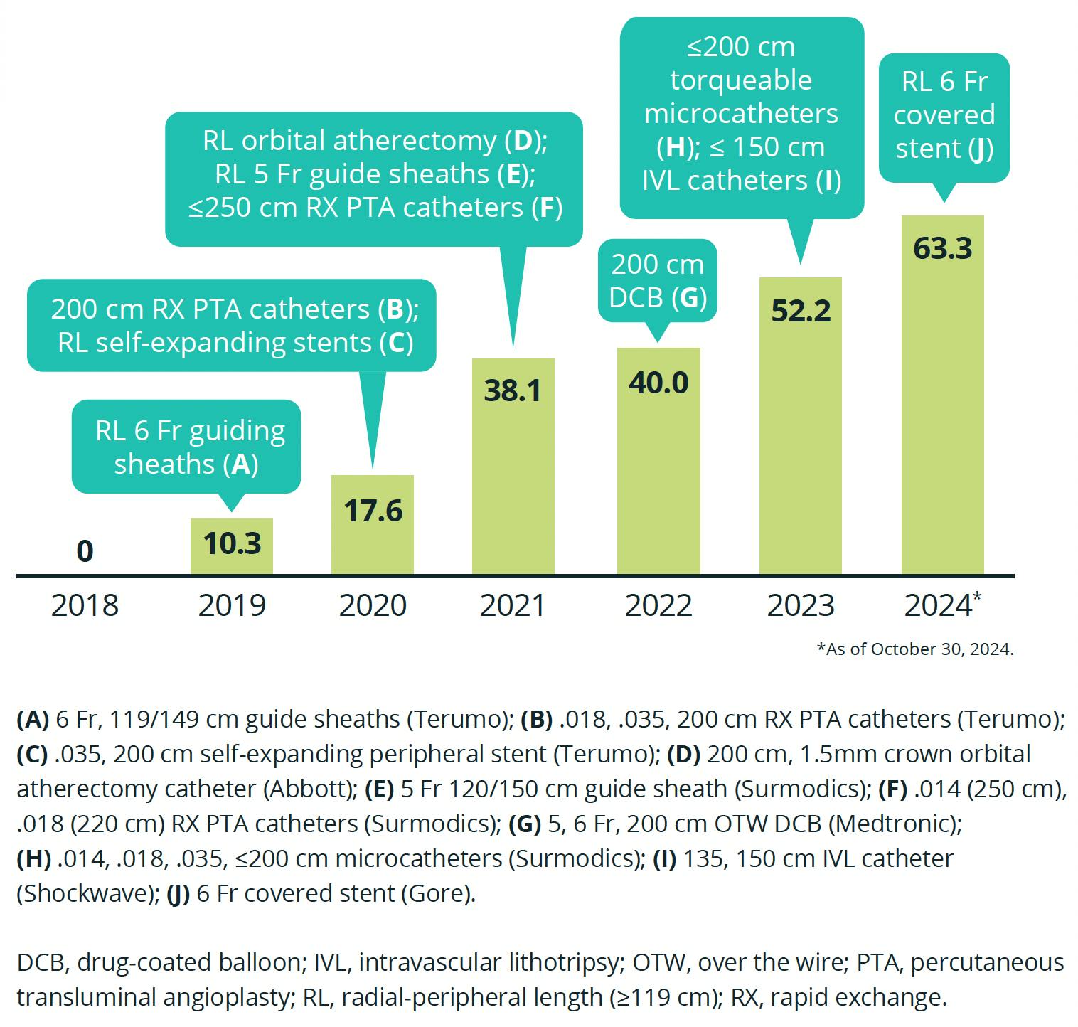
Figure 1. Progression of radial access utilization for peripheral interventions (%) with high-impact product adoptions by Sameh Sayfo, MD.
What other products have helped you increase the rate of peripheral interventions you perform from the radial approach?
Dr. Sayfo: The introduction of orbital and, later, laser atherectomy has been very helpful, specifically the Diamondback 360® orbital atherectomy system (Abbott) and Auryon™ atherectomy system (AngioDynamics, Inc.). The same with the IN.PACT™ 018, 200 cm drug-coated balloon (DCB; Medtronic). In the past, we needed to treat some younger patients via femoral access because we didn’t have radial-peripheral–length DCBs. In my opinion, it’s not fair to just balloon and stent given the great data we have with DCBs.13
Over the past few years, Surmodics has also introduced products that have made a huge impact on my practice. With the Sublime™ .014 and .018 RX PTA catheters [250 cm and 220 cm working lengths, respectively] (Surmodics, Inc.), we were able to do below-the-knee (BTK) work. Surmodics also introduced the Sublime™ microcatheters in 2023. That really changed the game, because now we have 200 cm microcatheters in .014, .018, and .035, and we’re able to navigate complicated cases such as chronic total occlusions (CTOs) in the SFA, popliteal artery, or BTK lesions. The Sublime™ 5 Fr guide sheath has also been very useful for patients with small arteries, especially older, thinner females. All of these products have made us comfortable treating more challenging cases.
Morgan, from your point of view as a scrub tech, has radial access impacted your workflow or the complexity of cases?
Ms. Weems: Not really. Even though radial access might involve learning an extra step or two, with repetition you get it down and it becomes second nature. There are technical differences, such as prepping pedal access as backup when we’re going radial access. But overall, radial procedures are now faster for us to perform than femoral procedures and allow for faster turnover. This potentially allows us to do more cases in a room per day.
Dr. Sayfo, as a physician with a master’s in business administration, what are your thoughts on the economics of radial versus femoral access for PAD?
Dr. Sayfo: Data from the coronary side show you can eliminate a tremendous amount of cost for patients, hospitals, and health care systems by increasing rates of same-day discharge with radial access.14 We just published a retrospective study of 143 radial-peripheral patients we treated between 2020 and 2022.15 Our rate of same-day discharge was 97%. And remember, we didn’t have a lot of the equipment back then that we have now, such as radial-peripheral–length DCBs or laser atherectomy. That’s what we’re trying to achieve—to send patients home the same day, safely. Same-day discharge is a driver of patient satisfaction. People always want to sleep in their own beds—nobody likes staying in a hospital.
We also need to combat the biggest risk associated with interventions: access site bleeding. Population trends are making this even more important. Age, BMI, comorbidities, and case complexity increase bleeding risk.8,16 Women have higher access site bleeding complications than men.17 You can minimize these risks by going radial access.18-20
Reducing complications with radial access also results in cost savings.21 With radial, even if you have an access site complication, you rarely need to take the patient to surgery to repair it.22 For groin access procedure complications, it’s not uncommon to have to resort to open surgery, blood transfusion, vasopressors, or thrombin injections, things requiring intensive care unit admission.23 There are so many things that can go wrong.
What advice would you offer physicians seeking to implement the radial-to-peripheral approach?
Dr. Sayfo: There are two partnerships you need to develop. The first is with your staff, and the second is with your administration. Let’s start with the staff.
If the staff has never done a radial case before, you’re going to meet resistance. They’re going to assume the procedures will take longer. You need to explain to the staff why you’re taking this approach and make them part of the solution. Educate them on the technology and what is expected of them. For example, if you’re going to use ultrasound every time—well, almost every vascular interventionalist uses ultrasound for groin access, so it’s not going to be an issue. Our staff has reached the point where they will text me before each case “radial, yes? Or radial/pedal?” When I reply “femoral,” their response is “really?” Radial has become the norm.
Morgan, any tips for staff who are new to radial-to-peripheral procedures?
Ms. Weems: It’s really important to make sure all the sheaths remain wet right up to the moment they’re inserted; otherwise, they can be painful for the patient. I always keep wet 4 X 4 inch sterile gauze pads over the sheaths until they’re needed, to keep them hydrophilic. I’ll also flush and prep the small sheath after it’s been used at the beginning of the procedure just in case it needs to be used again.
What kind of partnership do you need to develop with administration?
Dr. Sayfo: Well, at a basic level you’re going to need administration to support you in getting the equipment you’ll need. You can start with diagnostic procedures, which are easy to do with the equipment you already have. This applies to simple iliac cases using, say, 150 cm balloons, DCBs, or stents. But when you move on to SFA, popliteal, BTK, or complex cases, you really need to partner with someone in administration. I’ve been extremely fortunate to be able to partner with Ellie and the rest of the administration for this.
Ms. Huff: The key to successfully launching a new approach or program is for a physician, like Dr. Sayfo, to collaborate with someone like me in administration to develop an adoption strategy. We need to determine how to communicate the message and secure investment for the proposed program. This collaboration, which we call a dyad between an executive partner and a physician leader, is the secret sauce for success.
How does this collaboration work?
Ms. Huff: Physicians often lack the time to create detailed presentations or may not have training in change management. When introducing a new concept, you’ll inevitably encounter resistance. Therefore, you need a strategy to promote the idea, implement it, and monitor progress over time. An administrator typically has the time for these tasks, whereas physicians are usually occupied with patient care. Although physicians may have access to the latest approaches, tools, and innovations, they might not have the time to navigate the organization’s processes for acceptance and procurement.
Challenges can arise with supply chain management, but if you’ve already prepared a pitch deck with your administrator that outlines the implications and value proposition, you’re better equipped to handle them. Not everyone will be on board initially, but I’ve found that if I partner with early adopters and have a passionate physician leading the change, we’re more likely to succeed.
RADIAL-PERIPHERAL PRO TIPS
What’s your top tip for new radial operators?
Dr. Sayfo: As basic as it sounds, you really need to attend a course on radial access. After that, start with simple cases and make sure you have all the equipment you need—don’t go ad hoc (Figure 2). There’s nothing worse than being surprised without the right tools. I have a rule of four: (1) know your equipment, (2) know your staff, (3) know your skills, and (4) know your patients. The more experience you get with radial-to-peripheral, the more comfortable you become doing complex cases.
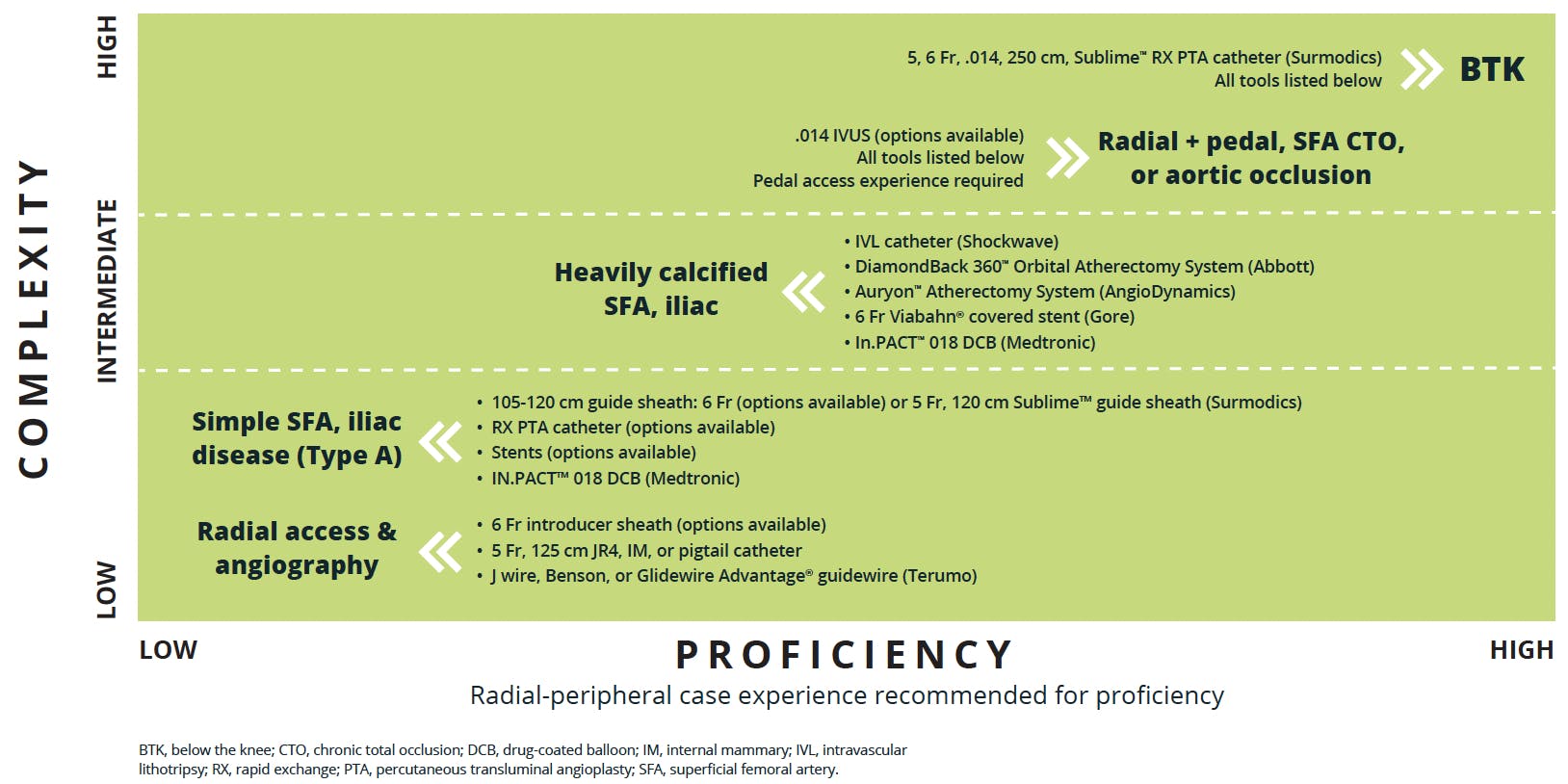
Figure 2. Radial-to-peripheral learning curve and recommended toolkit. Based on current practice and clinical experience of Sameh Sayfo, MD.
How often do you perform peripheral cases solely from the wrist, without additional access?
Dr. Sayfo: Most of my cases are radial alone. Of course, the groin is always prepped just in case, but our crossover rate to femoral is ≤ 3%.15 Whenever I have a patient with complex SFA anatomy, a CTO, or a previous stent that clearly looks undersized, I also prep pedal access. Sometimes I might want to complement a radial case with just a wire and snare from pedal access to get an arterial railway or go pedal with a sheath for combined radial/pedal therapy.
As with any other intervention, the more complicated the case, the more likely you are to need dual access. Surprisingly enough, I think one of the most complicated cases is aortoiliac occlusion, and I’ve done many of these cases with dual-radial or -ulnar access, no groin or pedal stick.
Do you prefer left or right radial?
Dr. Sayfo: I prefer right radial, mainly because our rooms are set up with the physician on the right side of the table. For a very small percentage of patients, I’ll choose the left radial when I expect navigation of the arch will be difficult or the patient is very tall, but most of the time that’s not a problem. The moment I have a long sheath where I want it, right or left doesn’t matter as much as it did in the past. The devices are much longer now, which reduces the need to go left radial to obtain more length.
Tell us a little more about how you set up for radial.
Dr. Sayfo: How you set up the table can be important. If you go left radial, and the patient’s arm is extended and you have two long tables, then the radiation protection is only protecting you. That’s one of the reasons I don’t choose the left radial. When I go with right radial, the staff can’t feel the difference between going femoral or radial. The monitors in your room may also be set up for working from the right side of the patient, making it uncomfortable to keep an eye on the monitors if you choose the left radial. So, the room should be set up in a way that’s comfortable for you, comfortable for the patient, and avoids you getting in the way of the staff.
For example, if you put a long table on the left side, you’re taking up a lot of the space in a cath lab. That’s the side where the nurses are standing, where the medications are for the nurses to administer, and where the devices and equipment are. The nurses won’t have enough room to bring you devices and you’re going to be in the way. Even when I go left radial, I don’t put in a long table or extend the patient’s arm—I just move the arm up and over, so I can work from the right side, or I stay on the left side but I move the arm to the side of the left groin and continue the case as if I had left groin access.
How do you obtain radial access?
Dr. Sayfo: I now use ultrasound to size the vessel 100% of the time. If the vessel is too small for a 6 Fr guide sheath, I might use the Sublime™ 5 Fr guide sheath. Alternatively, I might use the ulnar artery on the same [right] arm or the left radial artery if either of those are bigger.
Ultrasound-guided access is really important, especially for a physician who does not do a lot of radial cases. Ultrasound access saves time and reduces the number of times you have to do a stick—that’s clearly demonstrated in the literature.24 Using ultrasound guidance, we obtain radial access and insert a 6 F short sheath. We use a Terumo system, but there are others. Next, we always give a mixture of nitroglycerin and verapamil to help vasodilate the radial artery.
What is your radial cocktail?
Dr. Sayfo: We combine 400 μg nitroglycerin with 5 mg verapamil. Usually, I administer half the dose at the beginning of diagnostics, then we give about 50 U/kg of heparin through the peripheral intravenous line. Some operators mix heparin with the vasodilation cocktail, but we usually give it intravenously.
Which catheter do you use for imaging?
Dr. Sayfo: I usually use a JR4 5 Fr catheter for diagnostics—I like the small profile and long length. Sometimes we have patients who have had an EVAR or aortobifemoral bypass, which can take up length. A 125 cm JR4 catheter can get you anywhere you need for your diagnostic picture, no matter the patient’s height or anatomy. Also, since I usually go right radial, the JR4 curve helps me navigate the arch.
My number two choice is a 5 Fr, 125 cm internal mammary catheter, and my number three choice is a pigtail catheter. A pigtail catheter takes a little finesse to use, but it works well because you can flip it to the arch. If you’re going left radial with a pigtail, you just go over the wire; there’s not much need for manipulation. Some operators like a pigtail because it can be positioned to visualize the aortobifemoral bifurcation and aortic runoff, but I usually don’t perform a distal aortic angiogram unless I’m dealing with an aortobiiliac occlusion. If I’m dealing with SFA or any selective disease, I do selective pictures. This saves you contrast and reduces radiation.
What about wire selection?
Dr. Sayfo: I start with a table J wire. If that doesn’t cross the arch, then I switch to a Benson or a soft Glidewire® guidewire (Terumo Interventional Systems). When you’re using a Glidewire® guidewire, it’s very important to always keep an eye on the tip of the wire, because if you’re not paying attention it can go into the superior mesenteric or renal arteries and perforate. If that happens, you have a big problem on your hands before you’ve even started your case. I deliver my JR4 catheter into the common iliac or external iliac artery as distal as possible and we start with selective angiograms.
Next, if you’re going to convert to intervention, you’ll need to switch out your diagnostic catheter and sheath to a long radial-to-peripheral sheath, and for that it will be better to utilize a stiff wire like a Supra Core™ guidewire (Abbott).
How do you select your interventional sheath?
Dr. Sayfo: If I’m going to work on a common iliac or aortobiliac occlusion, I’ll typically use a 105 cm radial-to-peripheral sheath. For the external iliac, SFA, or popliteal arteries in an average-size patient, I usually use a 119 or 120 cm sheath. If the patient is 6 ft or taller or has had an aortobifemoral bypass or an EVAR, I might switch to a longer sheath, especially if I’m dealing with distal SFA, popliteal, or BTK disease.
How do you deliver the sheath?
Dr. Sayfo: For sheath delivery, I replace my J wire with a stiff wire through the JR4 catheter. As I said, my preference is a Supra Core™ guidewire, though some operators prefer Amplatz or Glidewire® guidewires. But again, if you use a Glidewire®, you need to keep an eye on the tip to avoid complication. After I place the stiff wire, I take out the JR4 and give my second dose of radial cocktail, heparin 50 U/kg, to reach full heparinization. My goal is an activated clotting time (ACT) of 250 seconds. Then I take out my radial access sheath and deliver the long sheath.
No matter what product you’re using, the sheath should be wetted with saline to activate the hydrophilic layer of the sheath. For successful delivery of the sheath, you need to ensure you have a nice rail with the stiff wire and make sure to just keep advancing the sheath—don’t pause halfway—until you get to the external iliac or wherever you want to land it. Otherwise, you might run into a spasm, and that makes it hard to push the sheath. Now I have the sheath in place, I have heparinization, and a couple minutes later I’m going to get an ACT.
What comes next?
Dr. Sayfo: That depends on the disease and how I plan to treat it. If I’m going to treat the common femoral, SFA, popliteal, or BTK arteries—really, anything but an iliac case—I’ll deliver a microcatheter as deep as possible and switch to a ViperWire Advance™ 475 cm guidewire (Abbott), which is the only long wire we have. I put it all the way into the foot, and now I can do whatever else I need to do: orbital atherectomy, laser atherectomy, intravascular lithotripsy (IVL), DCB, or just a regular balloon and stent.
When I’m finished with treatment, it’s time to remove the sheath. We used to do this by putting the dilator back, removing the .014 wire, putting in a .035 wire, removing the sheath, and closing with either a TR Band® (Terumo Interventional Systems) or other vascular band. More recently, I’ve simply been removing the sheath and putting the short sheath back in (the one we used at the beginning). You can only do this if the patient is heavily sedated, and you can’t do this over an .035 wire.
ANSWERS TO COMMON RADIAL-PERIPHERAL OBJECTIONS
One objection you hear to the radial-peripheral approach is the lack of a bailout option. How do you feel about that?
Dr. Sayfo: Yes, a lot of operators will not do an iliac case with radial access because of the risk of iliac perforations, which can be fatal, and the concern of not being able to deliver a bailout device via a 6 F sheath. I actually love to have that conversation, because I think that iliac perforations are easier to manage from the radial compared with the femoral approach.
If you’re going femoral, you’re usually up and over with a 6 Fr system. You balloon the contralateral external iliac artery, and boom, you have a rupture. In this situation, the first thing you do is inflate the balloon to tamponade the bleeding and try to get contralateral access. That’s easy.
If you’re treating from the ipsilateral side, after you tamponade the perforation you cannot safely change to an 8 Fr covered stent system from that same access site. So, operators go up and over from the other side, but now you need to get your wire through the balloon that’s already there. The contralateral side may also be occluded or severely diseased. You’re working as fast as you can, because the patient’s hemodynamics can deteriorate very quickly during an iliac perforation, and blood pressure can drop 100 mm Hg within a few minutes. It just becomes a hard situation.
With radial access, regardless of left or right radial, you just reinflate the balloon. Now you have all the time you need to get groin access with an 8 Fr system. Next, you can easily put the wire through, deflate the tamponade balloon, deploy the covered stent, and you’re done.
We’ve had two perforations in iliac cases, and I was much less stressed handling those via radial access than I would have been with femoral access. Granted, you’re not going to be able to deploy an 8 Fr covered stent from radial access, but you can easily tamponade with the balloon from radial access. That gives you all the time you need, all the safety margin, to get 8 Fr groin access when the patient’s stable. Alternatively, Gore & Associates now offers a 6 Fr compatible, balloon-expandable covered stent (VIABAHN® VBX balloon expandable endoprosthesis) that can be delivered via radial access.
What are some other common objections to radial-to-peripheral and your take on those?
Dr. Sayfo: Common objections include:
- Risk of stroke. There has been concern about the risk of stroke with radial access in general because you’re crossing the aortic arch. As others have reported,25 I don’t think the data support this concern. Our single-center study showed a < 1% stroke rate among 143 radial-peripheral patients.15 Another paper came out last year on the first prospective, multicenter registry on radial access for PAD.26 There were no reports of stroke among more than 100 patients. Remember, with radial access, the moment you have your catheter and stiff wire in and you place your sheath, you have a tube that’s delivering all the devices. There’s not a lot of motion in the arch while you’re working.
- Long learning curve. The idea that radial access requires a long learning curve depends on your background and the complexity of radial-peripheral procedures you want to do. It’s easy to teach a cardiologist to get started with radial-to-peripheral because they’ve already learned the access part of it, which is what matters the most. Good interventional radiologists have been using radial or brachial access for a long time for prostate artery embolization and gastrointestinal bleeds.
Even if you’ve never learned radial access, with ultrasound, radial access is not hard—it’s sometimes easier than femoral access. After you learn access, the main thing is learning navigation of the arch. If you start with diagnostics and simple iliac and SFA cases, it takes you about 10 to 30 cases to become comfortable. Doing diagnostic angiograms with radial access is faster and safer from radial compared with femoral access—no groin stick, no downtime, and the patient can go in for surgery the same day. After you’re comfortable doing that, you can move on to more complex iliac and SFA cases (Figure 2).
High-complexity cases involve SFA CTOs, distal aortic occlusions and, at the most complex level, BTK occlusions. For these you need to understand pedal access. Unfortunately, a lot of good operators either don’t do any pedal cases or very few, and you need experience to become proficient. Intravascular ultrasound (IVUS) is also extremely important in aortic occlusion cases because you need to know where you can reenter. You can use some reentry devices from the radial approach for the iliacs, but you need to understand how long they are and what sheath to use for them. For example, you cannot use anything longer than a 120 sheath because the reentry device has a 120 cm shaft length (Pioneer Plus™ IVUS-guided reentry catheter, Philips). Aside from that, in my view, when tackling a BTK lesion you really need to familiarize yourself with radial-to-peripheral length laser atherectomy and the Sublime™ 250 cm .014 RX PTA balloon.
- Longer procedure/fluoroscopy time. The data do not support prolonged procedure time and more radiation exposure for radial versus femoral access. We published an abstract at TCT 2023 that retrospectively compared iliac intervention via femoral access (n = 72) versus radial access (n = 72).27 There was no difference between the two groups in procedure time or volume of contrast media. In fact, the only difference between the groups was shorter length of stay for radial versus femoral (1.0 vs 1.3 days, respectively; P = .044) and a trend toward lower 1-year mortality for radial versus femoral (3% vs 11.1%; P = .067). Similar data have been published in the coronary literature.28
- Lack of devices. Another myth is that there are not enough radial-to-peripheral devices. I don’t think that’s accurate; a lot of operators simply don’t know what’s available (Figure 1). For example, many physicians don’t know there’s a 200 cm DCB (IN.PACT™ 018) or radial-to-peripheral length orbital and laser atherectomy devices (Diamondback 360® peripheral orbital atherectomy system and Auryon™ XL catheter, respectively). They also don’t know about the Sublime™ 200 cm microcatheter, which is a game-changer, and the Sublime™ 220 cm and 250 cm RX PTA balloons (.018 and .014 profiles, respectively). There are still some gaps, but they don’t prevent me from doing most of my peripheral cases from radial access.
What do you see as the principal remaining gaps in the radial-to-peripheral toolkit?
Dr. Sayfo: The Achilles’ heel for radial-to-peripheral has been lack of a long wire. We use the ViperWire Advance™ 475 cm guidewire (Abbott), but I’m not satisfied with the support it provides. Having said that, we just did a case involving an old, occluded stent from the popliteal down to the tibioperoneal trunk (TPT) and peroneal artery. We crossed it with the Sublime™ microcatheter and wire escalation, put in the 475 cm wire, performed laser atherectomy, ballooned, and used the DCB. That’s exactly what everybody else would have done from femoral access.
Lack of radial-peripheral–length IVUS remains a limitation, and there’s a limitation for bioresorbable stents. The Esprit™ BTK resorbable scaffold system (Abbott) is now commercially available, but that’s a 145 cm working length. So those are challenges for BTK.
I would also like to see more stent options. We have stents from Terumo Interventional Systems (R2P™ MISAGO® RX self-expanding peripheral stent) and Cordis (SMART Radianz™ vascular stent system). I really like the stent from Terumo Interventional Systems, but it’s a .035 system. Sometimes I’ll already be working with an .014 wire, so it can be challenging to deliver an .035 stent on an .014 system. But the balloon catheters, microcatheter, and guide sheaths that Surmodics offers are the most important things. We just need a good radial-length wire, more stent options, and IVUS to complete the package.
EXPERIENCE WITH SUBLIME™ RADIAL ACCESS PRODUCTS
Let’s dig a little deeper into your experience with the Sublime™ Radial Access Platform, beginning with the guide sheaths.
Dr. Sayfo: The fact that Surmodics offers the Sublime™ guide sheath in 5 Fr as well as 6 Fr has been very helpful for patients with small arteries. This sheath is very trackable and deliverable, which is great, because you’re always worried about tracking a stiff wire through the tortuosity of the brachiocephalic artery and the aortic arch. This sheath tracks a stiff wire very easily, and when the sheath is settled down where you want it, let’s say in the common or external iliac artery, you don’t have to worry about it moving. This gives you the stability you need to take care of the patient without worrying about the sheath pulling back and losing that support.
Do you see advantages in using Sublime™ PTA catheters?
Dr. Sayfo: The balloons have been great, and they have different advantages. For one, they have a variety of profiles. The .014 has a working length of 250 cm, which allows you to reach the BTK arteries. Their performance is also important. The first case I performed with a Sublime™ PTA catheter was a complete SFA CTO, and I was worried the balloon was not going to cross, but it crossed very easily. The rapid-exchange design of these catheters makes handling them much easier. For example, if I cross with a .018 and I want to use just a balloon, I don’t have to switch to a long wire.
How about the Sublime™ microcatheters?
Dr. Sayfo: The 200 cm length of these microcatheters is extremely important, but the design is also very good. These microcatheters are very torqueable and deliverable. Most of the time I use an angled tip, which is very important in CTO cases, where there is a higher chance you’ll need to perform a dissection reentry technique. An angled tip catheter allows you to get back to the true lumen distally.
It’s also useful to have the Sublime™ .014, .018, and .035 microcatheters. Sometimes in a calcified or tortuous artery, or a distal artery, I like to use either the .018 or .014 microcatheters with their smaller profiles so I can cross farther. These are also very, very slippery catheters. I can deliver these through a 100% occlusion without any issues.
What kinds of procedures do Sublime™ microcatheters help enable?
Dr. Sayfo: It can be really hard to deliver a .014 wire, such as the 475 cm ViperWire Advance® (Abbott) guidewire, through a lesion unless its only, for example, a 70% occlusion that anything can cross. If you have a highly calcified, tortuous lesion or a CTO, you’re going to have to cross with a hydrophilic wire, such as a Glidewire® or Hi-Torque Command™ (Abbott) guidewire. Then, after you cross with that, you want the microcatheter to track through that wire and serve as a delivery vehicle that allows you to put the 475 cm ViperWire Advance® guidewire where you want it.
At this point I use a Sublime™ microcatheter for most of my cases, unless I’m dealing with a proximal iliac that doesn’t require the 200 cm length or a 70%-type lesion where I can cross with the ViperWire Advance® guidewire by itself.
Caution: Federal (US) law restricts the Sublime™ Radial Access Guide Sheath, the Sublime™ Radial Access .014 and .018 RX PTA Dilatation Catheters, and the Sublime™ Radial Access .014, .018, and .035 Microcatheters to sale by or on the order of a physician. Please refer to each product’s Instructions for Use for indications, contraindications, warnings, and precautions. SURMODICS, SUBLIME, and SURMODICS and SUBLIME logos are trademarks of Surmodics, Inc. and/or its affiliates. Third-party trademarks are the property of their respective owners.
1. Vascular Verification Program Verifies First Four Hospitals. News release. American College of Surgeons. June 14, 2023. Accessed Oct. 18, 2024. https://www.facs.org/media-center/press-releases/2023/vascular-verification-program-verifies-first-four-hospitals/
2. Mason PJ, Shah B, Tamis-Holland JE, et al. An update on radial artery access and best practices for transradial coronary angiography and intervention in acute coronary syndrome: a scientific statement from the American Heart Association. Circ Cardiovasc Interv. 2018;11:e000035. doi: 10.1161/HCV.0000000000000035
3. Meertens MM, Ng E, Loh SE, et al. Transradial approach for aortoiliac and femoropopliteal interventions: a systematic review and meta-analysis. J Endovasc Ther. 2018;25:599-607. doi: 10.1177/1526602818792854
4. Liu LB, Cedillo MA, Bishay V, et al. Patient experience and preference in transradial versus transfemoral access during transarterial radioembolization: a randomized single-center trial. J Vasc Interv Radiol. 2019;30:414-420. doi: 10.1016/j.jvir.2018.10.005
5. Criqui MH, Matsushita K, Aboyans V, et al. Lower extremity peripheral artery disease: contemporary epidemiology, management gaps, and future directions: a scientific statement from the American Heart Association. Circulation. 2021;144:e171-e191. doi: 10.1161/CIR.0000000000001005
6. Vemulapalli S, Greiner MA, Jones WS, et al. Peripheral arterial testing before lower extremity amputation among Medicare beneficiaries, 2000 to 2010. Circulation Cardiovasc Qual Outcomes. 2014;7:142-150. doi: 10.1161/CIRCOUTCOMES.113.000376
7. Trust for America’s Health. The state of obesity 2020: better policies for a healthier America. Accessed October 14, 2024. https://www.tfah.org/report-details/state-of-obesity-2020/
8. Benamer H, Louvard Y, Sanmartin M, et al. A multicentre comparison of transradial and transfemoral approaches for coronary angiography and PTCA in obese patients: the TROP registry. EuroIntervention. 2007;3:327-332. doi: 10.4244/eijv3i3a60
9. Cooper CJ, El-Shiekh RA, Cohen DJ, et al. Effect of transradial access on quality of life and cost of cardiac catheterization: a randomized comparison. Am Heart J. 1999;138:430-436. doi: 10.1016/s0002-8703(99)70143-2
10. Lentz CM, Zogaj D, Wessel HK, et al. Brachial and axillary artery vascular access for endovascular interventions. Ann Vasc Surg. 2022;81:292-299. doi: 10.1016/j.avsg.2021.09.052
11. Posham R, Biederman DM, Patel RS, et al. Transradial approach for noncoronary interventions: a single-center review of safety and feasibility in the first 1,500 cases. J Vasc Interv Radiol. 2016;27:159-166. doi: 10.1016/j.jvir.2015.10.026
12. Bazan HA, Le L, Donovan M, et al. Retrograde pedal access for patients with critical limb ischemia. J Vasc Surg. 2014;60:375-382. doi: 10.1016/j.jvs.2014.02.038
13. Laird JA, Schneider PA, Jaff MR, et al. Long-term clinical effectiveness of a drug-coated balloon for the treatment of femoropopliteal lesions. Circ Cardiovasc Interv. 2019;12:e007702. doi: 10.1161/CIRCINTERVENTIONS.118.007702
14. Lindner SM, McNeely CA, Amin AP. The value of transradial: Impact on patient satisfaction and healthcare economics. Interv Cardiol Clin. 2020;9:107-115. doi: 10.1016/j.iccl.2019.08.004
15. Sayfo S, Salih M, Moubarak G, et al. Safety and efficacy of radial artery access for peripheral vascular intervention: a single-center experience. Am J Cardiol. 2024;226:59-64. doi: 10.1016/j.amjcard.2024.06.025
16. Visonà A, Zurlo C, Panzavolta C, et al. Bleeding risk in patients with peripheral arterial disease. Life (Basel). 2022;13:47. doi: 10.3390/life13010047
17. Hess CN, Rao SV, Kong DF, et al. Embedding a randomized clinical trial into an ongoing registry infrastructure: unique opportunities for efficiency in design of the Study of Access site For Enhancement of Percutaneous Coronary Intervention for Women (SAFE-PCI for Women). Am Heart J. 2013;166:421-428. doi: 10.1016/j.ahj.2013.06.013
18. Cantor WJ, Mehta SR, Yuan F, et al. Radial versus femoral access for elderly patients with acute coronary syndrome undergoing coronary angiography and intervention: insights from the RIVAL trial. Am Heart J. 2015;170:880-886. doi: 10.1016/j.ahj.2015.08.011
19. Pandie S, Mehta SR, Cantor WJ, et al. Radial versus femoral access for coronary angiography/intervention in women with acute coronary syndromes: insights from the RIVAL trial (Radial Vs femorAL access for coronary intervention). JACC Cardiovasc Interv. 2015;8:505-512. doi: 10.1016/j.jcin.2014.11.017
20. Cox N, Resnic FS, Popma JJ, et al. Comparison of the risk of vascular complications associated with femoral and radial access coronary catheterization procedures in obese versus nonobese patients. Am J Cardiol. 2004;94:1174-1177. doi: 10.1016/j.amjcard.2004.07.088
21. Mitchell MD, Hong JA, Lee BY, et al. Systematic review and cost–benefit analysis of radial artery access for coronary angiography and intervention. Circ Cardiovasc Qual Outcomes. 2012;5:454-462. doi: 10.1161/CIRCOUTCOMES.112.965269
22. Alkagiet S, Petroglou D, Nikas DN, Kolettis TM. Access-site complications of the transradial approach: rare but still there. Curr Cardiol Rev. 2021;17:279-293. doi: 10.2174/1573403X16999200819101923
23. Ortiz D, Jahangir A, Singh M, et al. Access site complications after peripheral vascular interventions: incidence, predictors, and outcomes. Circ Cardiovasc Interv. 2014;7:821-828. doi: 10.1161/CIRCINTERVENTIONS.114.001306
24. Nguyen P, Makris A, Hennessy A. Standard versus ultrasound-guided radial and femoral access in coronary angiography and intervention (SURF): a randomised controlled trial. EuroIntervention. 2019;15:e522-e530. doi: 10.4244/EIJ-D-19-00336
25. Iezzi R, Posa A, Bilhim T, Guimaraes M. Most common misconceptions about transradial approach in interventional radiology: results from an international survey. Diagn Interv Radiol. 2021;27:649. doi: 10.5152/dir.2021.20256
26. Castro-Dominguez Y, Li J, Lodha A, et al. Prospective, multicenter registry to assess safety and efficacy of radial access for peripheral artery interventions. J Soc Cardiovasc Angiogr Interv. 2023;2:101107. doi: 10.1016/j.jscai.2023.101107
27. Sayfo S. Transradial versus transfemoral approach in peripheral vascular interventions. Presented at Transcatheter Cardiovascular Therapeutics 2023; October 23-26, 2023; San Francisco, California.
28. Mohandes M, Rojas S, Guarinos J, et al. Procedure time comparison between radial versus femoral access in ST-segment elevation acute myocardial infarction patients undergoing emergent percutaneous coronary intervention: a meta-analysis of controlled randomized trials. Heart Views. 2018;19:1-7. doi: 10.4103/HEARTVIEWS.HEARTVIEWS_82_16



Case Report
Successful Revascularization of SFA Stenosis Via Radial Access
By Sameh Sayfo, MD, MBA, FSCAI, FACC
Patient Presentation
A woman in her early 70s with a medical history of diabetes, hypertension, and hyperlipidemia presented to the hospital with worsening left lower extremity claudication (Rutherford class 3).
Diagnostic Findings
Arterial ultrasound and CTA showed a heavily calcified proximal left superficial femoral artery (SFA) stenosis. The plan was to revascularize this vessel utilizing a radial approach.
Treatment
Access was achieved through the patient’s right radial artery using ultrasound guidance and a radial cocktail was delivered. The radial access introducer sheath was exchanged for a 6 Fr, 120 cm Sublime™ guide sheath (Surmodics, Inc.), which was navigated to the stenosis. An angiogram confirmed that the patient had a heavily calcified proximal left SFA stenosis (Figure 1). A Hi-Torque Command™ 18 guidewire (Abbott) and 150 cm Navicross® support catheter (Terumo Interventional Systems) were used in conjunction to cross the lesion. The Hi-Torque Command™ 18 guidewire was swapped out for a .014, 475 cm ViperWire™ guidewire (Abbott).
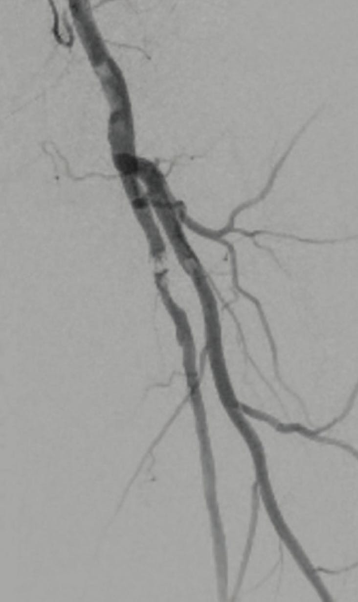
Figure 1. Initial angiogram displaying calcified left SFA stenosis.
Orbital atherectomy was performed using the Diamondback 360® peripheral orbital atherectomy system (Abbott) with a 1.75 mm solid crown (Figure 2). Following the atherectomy passes, a 5 X 60 mm IN.PACT™ 018 drug-coated balloon (Medtronic) was dilated across the lesion. The final angiogram showed 0% residual stenosis, patent flow, and no dissections (Figure 3).
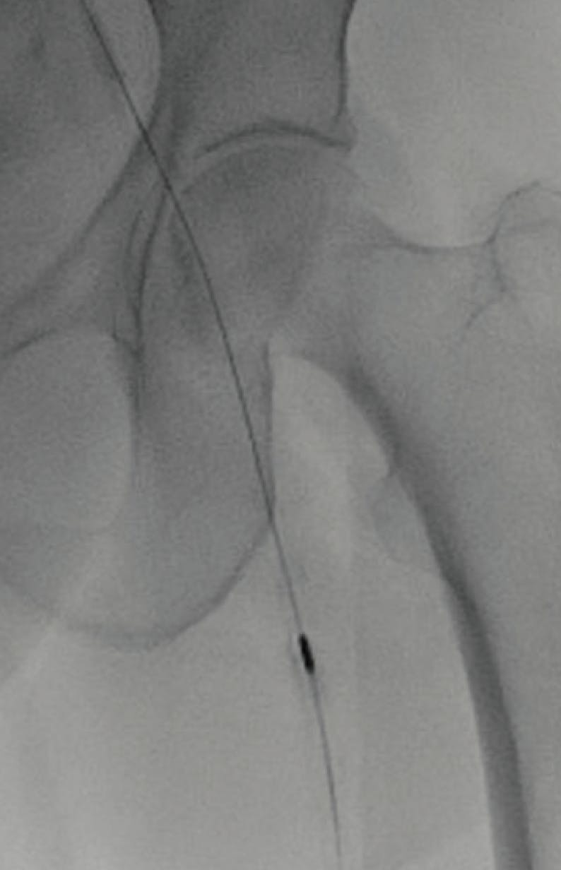
Figure 2. Orbital atherectomy was performed using the Diamondback 360® peripheral orbital atherectomy system with a 1.75 mm solid crown.
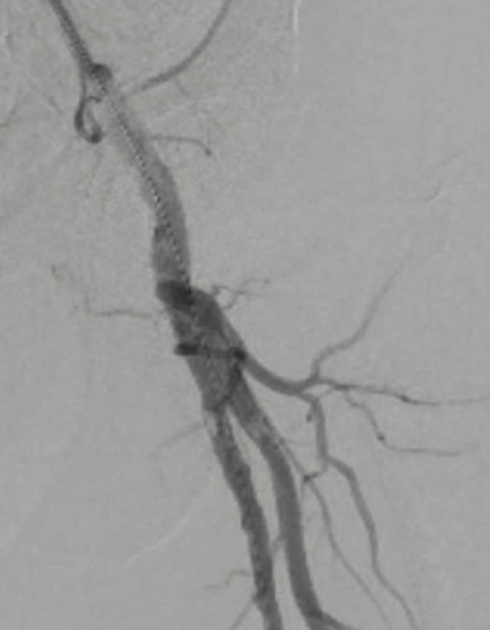
Figure 3. Final angiogram showed 0% residual stenosis, patent flow, and no dissections.
PostProcedure Outcome
The patient was discharged the same day with a complete resolution of her symptoms.
Caution: Federal (US) law restricts the Sublime™ Radial Access Guide Sheath, the Sublime™ Radial Access .014 and .018 RX PTA Dilatation Catheters, and the Sublime™ Radial Access .014, .018, and .035 Microcatheters to sale by or on the order of a physician. Please refer to each product’s Instructions for Use for indications, contraindications, warnings, and precautions. SURMODICS, SUBLIME, and SURMODICS and SUBLIME logos are trademarks of Surmodics, Inc. and/or its affiliates. Third-party trademarks are the property of their respective owners.
Case Report
Successful Revascularization of Aortoiliac Stenosis Via Radial Access
By Sameh Sayfo, MD, MBA, FSCAI, FACC
Patient Presentation
A woman in her mid 50s with a medical history of coronary artery disease, coronary artery bypass grafting, and diabetes presented with severe bilateral hip claudication.
Diagnostic Findings
Preprocedure ankle-brachial index (ABI) showed abnormality with readings of 0.23 on the right leg and 0.37 on the left leg (Figure 1). Follow-up CTA showed a distal aorta and bilateral common iliac artery occlusion (Figure 2).
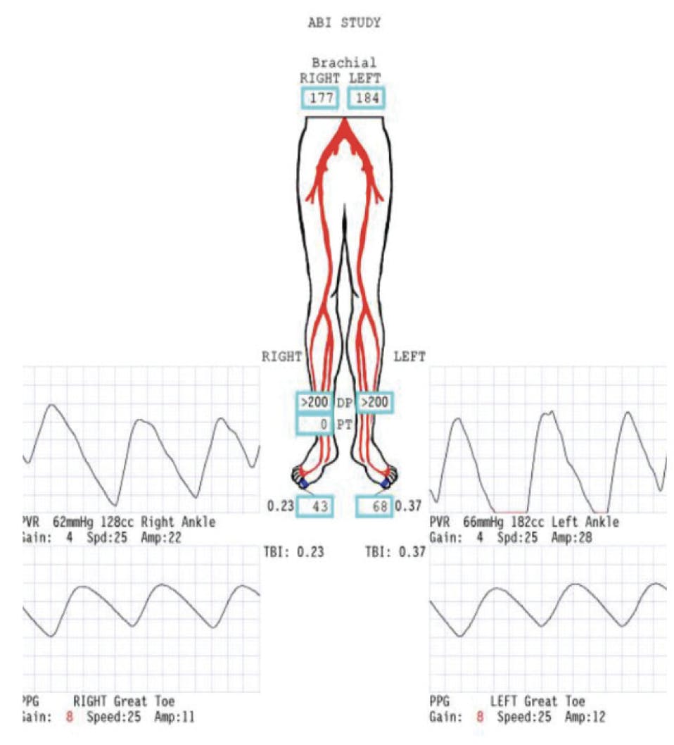
Figure 1. The patient’s initial ABI measurements.
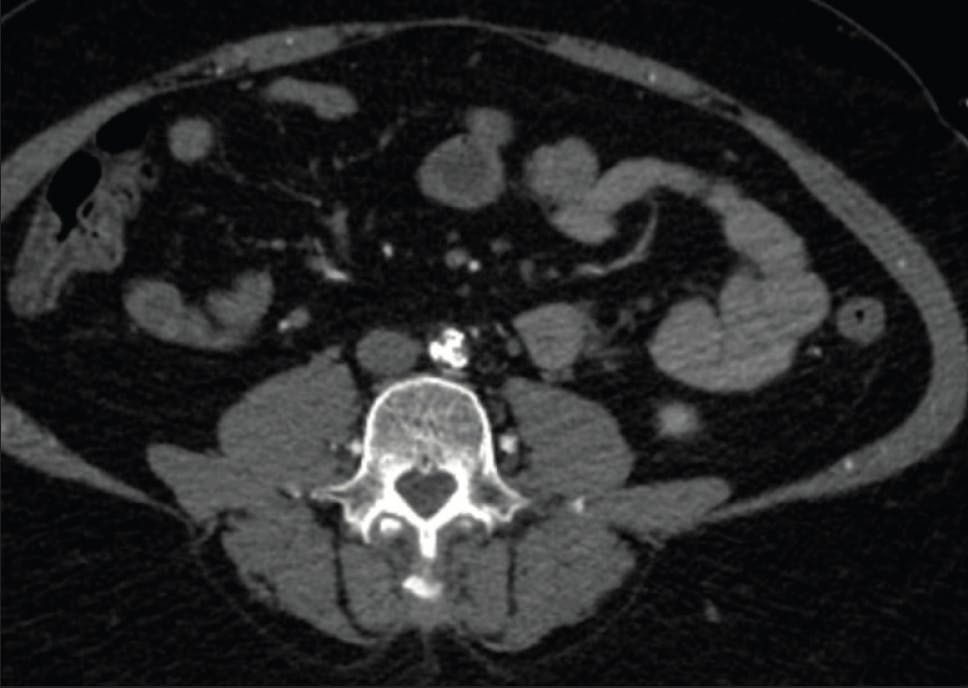
Figure 2. CTA showed a distal aorta and bilateral common iliac artery occlusion.
Treatment
Bilateral ulnar access was obtained using ultrasound guidance and radial cocktails were delivered. Follow-up angiography confirmed the prior CTA results. The two introducer sheaths were exchanged for two 6 Fr, 120 cm Sublime™ guide sheaths (Surmodics, Inc.), which were advanced to the distal aorta and into each occluded common iliac artery. Utilizing an .018 Glidewire Advantage® peripheral guidewire (Terumo Interventional Systems) and .035, 135 cm Navicross® support catheter through both sheaths, both lesions were successfully crossed. A 6 X 60 mm Shockwave™ M5+ intravascular lithotripsy catheter (Shockwave Medical) was used in both common iliac arteries to debulk the calcium. Two 8 X 80 mm R2P™ Misago® RX self-expanding peripheral stents (Terumo Interventional Systems) were deployed into each vessel, starting from the external iliac artery down to the distal common iliac artery. Additionally, two 7 X 39 mm RX AccuLink™ carotid stent system stents (Abbott) were deployed from common iliac to the distal aorta. The final angiogram showed complete resolution of the lesion, and the patient’s pain was also observed to be completely resolved the same day (Figure 3).
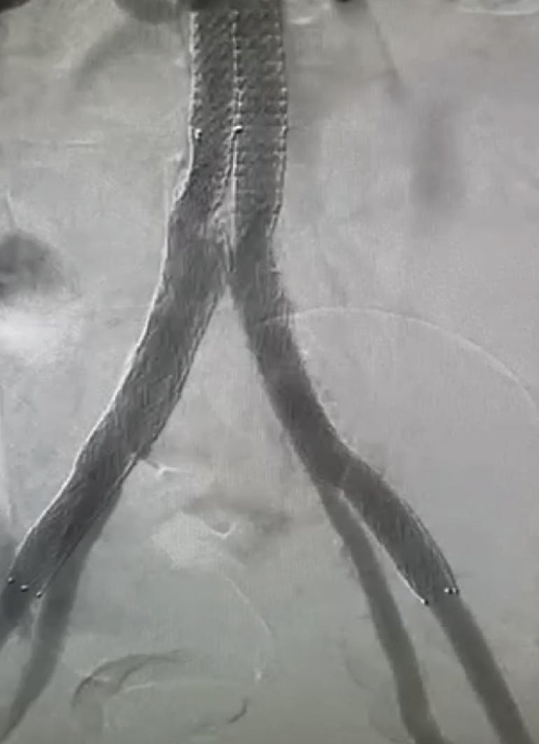
Figure 3. The final angiogram showed complete resolution of the lesion.
PostProcedure Outcome
The patient was discharged hours after the intervention.
Caution: Federal (US) law restricts the Sublime™ Radial Access Guide Sheath, the Sublime™ Radial Access .014 and .018 RX PTA Dilatation Catheters, and the Sublime™ Radial Access .014, .018, and .035 Microcatheters to sale by or on the order of a physician. Please refer to each product’s Instructions for Use for indications, contraindications, warnings, and precautions. SURMODICS, SUBLIME, and SURMODICS and SUBLIME logos are trademarks of Surmodics, Inc. and/or its affiliates. Third-party trademarks are the property of their respective owners.
Case Report
Successful Revascularization of BTK In-Stent Restenosis Via Radial Access
By Sameh Sayfo, MD, MBA, FSCAI, FACC
Patient Presentation
A man in his early 80s with extensive previous peripheral artery disease presented with resting pain. He had a previously placed stent extending from the peroneal artery to the distal popliteal artery. The patient had experienced contralateral groin access complications (pseudoaneurysm) in the past requiring thrombin injection.
Diagnostic Findings
Right radial access was achieved and a radial cocktail was delivered. The introducer sheath was exchanged for a 6 Fr, 150 cm Sublime™ guide sheath (Surmodics, Inc.). An initial angiogram showed an occluded distal popliteal and peroneal arteries with 100% restenosis of the previously deployed stent (Figure 1).
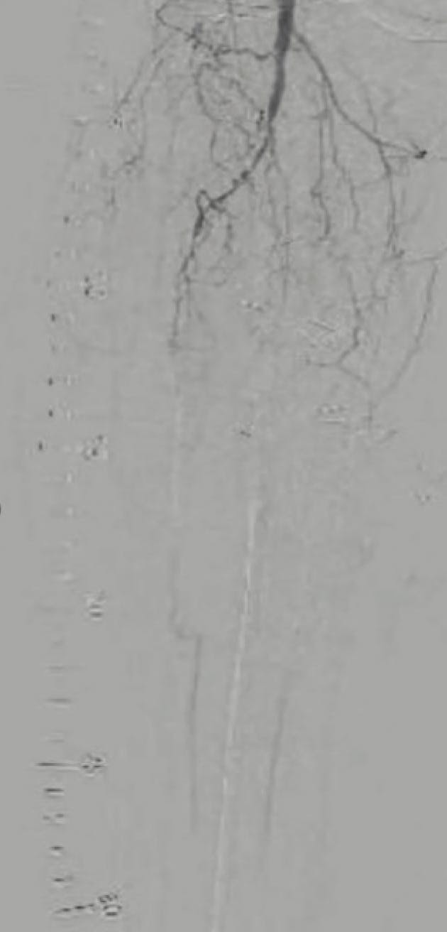
Figure 1. Initial angiogram showed occluded distal popliteal and peroneal arteries with 100% restenosis of the previously deployed stent.
Treatment
The Sublime™ guide sheath was advanced further into the vasculature. A 200 cm Sublime™ .018 microcatheter (Surmodics, Inc.) was introduced over the already placed .018 Glidewire Advantage® peripheral guidewire (Terumo Interventional Systems). The .018 Glidewire Advantage® guidewire was then swapped out for an .014 Astato® XS 20 guidewire (Abbott), and the lesion was then crossed. The Sublime™ microcatheter was removed, and the Astato® XS 20 guidewire was swapped out for a .014, 475 cm ViperWire Advance™ guidewire (Abbott).
Laser atherectomy using the Auryon™ 1.5 XL laser (AngioDynamics, Inc.) was performed throughout the stenotic segment. Next, an IN.PACT™ 018 drug-coated balloon (DCB; Medtronic) was dilated inside the stent (Figure 2). The DCB was removed, and a 2.5 X 100 mm, 250 cm, .014 Sublime™ Radial Access RX PTA catheter was advanced and dilated in the distal portion of the vessel for a final dilation.
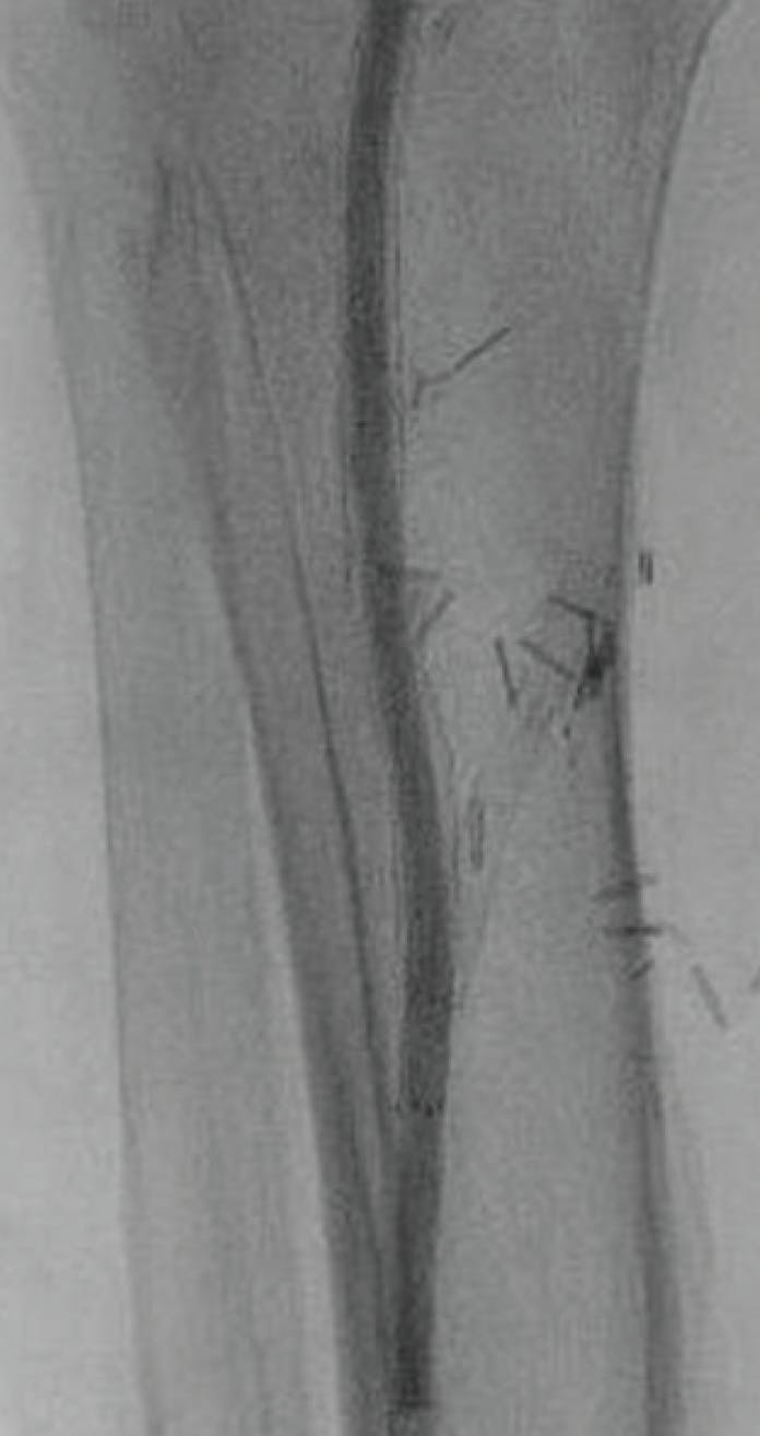
Figure 2. An IN.PACT™ 018 DCB dilated inside the stent.
A final angiogram showed single-vessel runoff to the plantar arch (Figure 3).
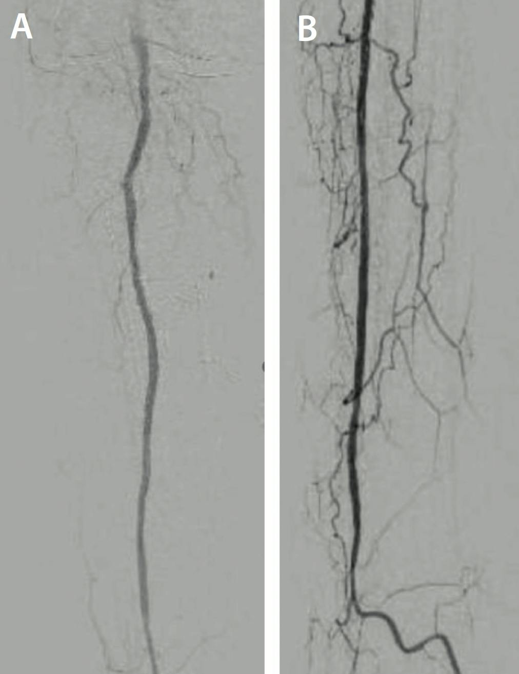
Figure 3. Final angiogram showed single-vessel runoff from distal popliteal artery (A) to the plantar arch (B).
PostProcedure Outcome
The patient was sent home the same day. At his 2-week follow-up, he had no pain.
Caution: Federal (US) law restricts the Sublime™ Radial Access Guide Sheath, the Sublime™ Radial Access .014 and .018 RX PTA Dilatation Catheters, and the Sublime™ Radial Access .014, .018, and .035 Microcatheters to sale by or on the order of a physician. Please refer to each product’s Instructions for Use for indications, contraindications, warnings, and precautions. SURMODICS, SUBLIME, and SURMODICS and SUBLIME logos are trademarks of Surmodics, Inc. and/or its affiliates. Third-party trademarks are the property of their respective owners.
Advertisement
Advertisement