Advertisement
Advertisement
August 2026
Sponsored by Boston Scientific Corporation
SEISMIQ™ IVL System as a Next-Generation Technology Offering for Complex PAD
Experts share their experience with SEISMIQ™ IVL and the differentiated device features that are expanding treatment possibilities for complex calcified lesions.
With Jaafer Golzar, MD, FACC, FSCAI, and Gregory A. Stanley, MD, FACS

The SEISMIQ™ Intravascular Lithotripsy (IVL) System (Figure 1) represents the next generation of IVL, purpose-built to address complex calcified lesions with maximum impact and precision. Combining advanced deliverability, enhanced visibility, and more consistent acoustic pressure, SEISMIQ is designed to help physicians maximize the impact of every pulse. Every aspect of the system has been thoughtfully engineered to provide greater control—from navigating challenging anatomies to delivering targeted therapy with clarity and precision.
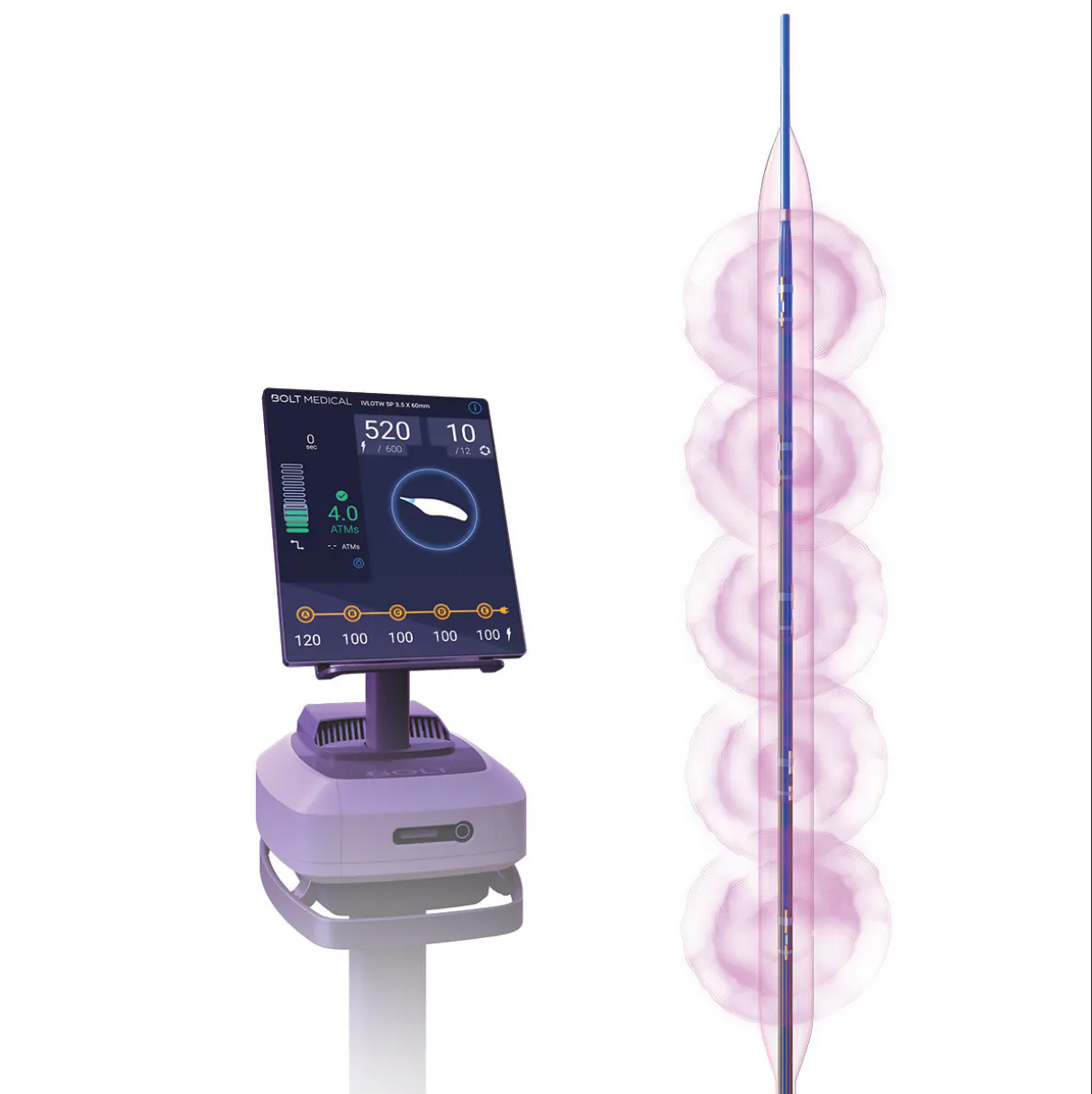
Figure 1. The SEISMIQ IVL System.
SEISMIQ IVL became commercially available in late 2025 and has been evaluated in the RESTORE ATK1 and RESTORE BTK2 clinical studies, both demonstrating 100% procedural success, 100% freedom from major adverse events at 30 days, low residual stenosis, and minimal use of adjunctive therapies.
As early adopters of this novel IVL system, Dr. Jaafer Golzar and Dr. Gregory Stanley of Advocate Health have gained firsthand experience incorporating SEISMIQ into clinical practice. In this feature, they share their perspectives on the technology, practical treatment algorithms, and case-based insights that highlight the differentiated features that have elevated the care of patients with peripheral artery disease (PAD).
From your clinical perspective, what’s driving the significant growth in IVL use over the past few years? What does IVL uniquely offer compared with other vessel preparation modalities available today?
Dr. Stanley: The significant growth of IVL use over the last few years is multifactorial but largely based in its ability to properly address a dominant and problematic obstacle in the peripheral vascular arena: moderate-to-severely calcified plaque. The percentage of patients with heavily calcified PAD is increasing dramatically due to an aging population with a higher prevalence of longstanding diabetes mellitus and chronic kidney disease. For those patients, standard interventional tools such as balloon percutaneous transluminal angioplasty (PTA), stenting, and atherectomy have noteworthy shortcomings and safety concerns, such as vessel recoil, flow-limiting dissection, perforation, distal embolization, inadequate lumen gain, and poor stent expansion, all of which contribute to worse procedural and long-term outcomes.
IVL technology is designed specifically to deliver energy waves for targeted calcium modification and normalization of vessel wall compliance, a mechanism that has proven effective in achieving acute lumen gain with a low-risk profile. With the known benefits of drug-elution technology,3-7 this vessel preparation strategy is particularly attractive for patients presenting with a heavy calcium burden.
The efficacy of this approach was established in the DISRUPT PAD III randomized controlled trial (and supporting observational data in nearly 1,400 real-world patients),8-10 which compared the use of IVL to PTA for vessel preparation prior to treatment with drug-coated balloon (DCB) angioplasty (with provisional stenting). In this trial, IVL provided superior procedural technical success, including residual stenosis (P < .0065), fewer flow-limiting dissections (3.5% vs 15.1%; P < .003), lower bailout stenting rate (4.6% vs 18.3%; P < .001), and higher primary patency at 1-year (80.8% vs 70.9%; P < .005) and 2-year follow-up (74.4% vs 57.7%; P < .005) versus PTA. This evidence added momentum to a fundamental shift in practice already underway, toward proactive calcium modification rather than reactive bailout strategies. That mindset positioned IVL to support a “leave-nothing-behind” approach in appropriate circumstances, while also enabling safer stenting when required. The final pieces encouraging IVL adoption are its ease of use, integration with intravascular ultrasound (IVUS) to augment standard angiography, and a low complication rate.
When considering a vessel prep strategy for complex PAD interventions with moderate-to-severe calcific lesions, there is a trade-off between treatment modalities in terms of efficacy, safety, efficiency, and durability. IVL offers a means to address calcium in all its forms: intimal, medial, adventitial, eccentric, and circumferential; and, it stands in contrast to atherectomy, which may excel in one calcium category but not another. The low-pressure mechanism is generally gentler than other modalities that may induce high-pressure barotrauma or more aggressive ablative methods. The balloon catheter is familiar to use with a minimal learning curve and supports a predictable and efficient workflow. Compared to other vessel prep options, IVL should be a front-line tool for patients with a high calcific burden, as it raises the standard for adequate vessel preparation.
As a recognized expert in IVL, how would you describe what SEISMIQ™ IVL (Boston Scientific Corporation) (Figure 1) brings to the table that other IVL technologies do not?
Dr. Golzar: As a long-time user of IVL for complex, heavily calcified lesions, I was initially skeptical that a new platform could offer a meaningful clinical advantage over established legacy technologies. However, evaluating the SEISMIQ system revealed four major advantages that directly optimize complex lesion management:
- Pulse capacity: The system provides up to 600 pulses per balloon, significantly exceeding legacy peripheral IVL capacities and allowing for the efficient treatment of multi-level or long-segment calcific disease without standard device exchanges.
- Acoustic mechanism: The system uses a laser-based energy source delivered via optical fibers to create a high-energy plasma plume, which subsequently expands into a cavitation bubble. This mechanism generates consistent, concentrated acoustic waves producing superficial and deep calcium disruption, fracturing the rigid calcific plaque.
- Pulse conservation and emitter selectivity: We can also select individual treatment segments and isolate specific emitters, which actively conserves our total pulse count for where it is needed most.
- User interface and workflow: The slick, highly intuitive touchscreen user interface is exceptionally user-friendly. Combined with advanced catheter deliverability and a fully sterile setup that completely eliminates handle-bagging, these operational and structural advantages have fundamentally shifted my clinical practice from legacy platforms to the SEISMIQ IVL system.
Dr. Stanley: SEISMIQ represents the next generation of IVL devices on the market, not simply the next iteration of IVL. Its multiple technologic advancements and updated design features take the best of what IVL is and make it better, while also addressing some pain points of currently available devices. SEISMIQ has changed how I think about using IVL and offers a new level of control for calcium modification that did not previously exist.
The laser-based pulses are strong, effective, and for the first time, directional. Each emitter station is easily visualized and is offset by 72° (Figure 2), which allows for both circumferential treatment along the length of the lesion, as well as for concentrated directed treatment at a recalcitrant lesion.
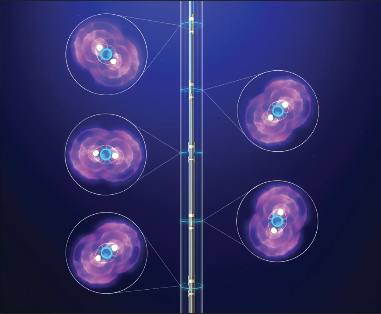
Figure 2. SEISMIQ IVL balloon catheter illustrating emitter station orientation—each offset by 72°—that enables directional, targeted therapy.
Another new and unique feature that has really impacted my ability to understand what is occurring during IVL treatment is the console displaying real-time balloon inflation pressure and pressure differential data. This readily accessible data helps to track the progress of treatment at a specific lesion segment and may ultimately prove to be a critical clinical indicator for adequate calcium modification.
My experience thus far with SEISMIQ IVL has instilled confidence in treating nearly all forms of calcific plaque, but especially eccentric and nodular lesions.
How can these features uniquely support fracture of eccentric and nodular lesion morphologies?
Dr. Stanley: The ability to fracture eccentric and nodular calcific plaque amounts to a balance of adequate energy delivery to the lesion without overtly destructive damage to adjacent tissue. We do not yet have the data to predict exactly how much energy delivery is required for each specific lesion—likely to be a product of calcium thickness, volume, and composition—but, theoretically, all lesions have a threshold of energy at which fracture is assured. We are accordingly left with a scenario not dissimilar from using a wrecking ball to demolish a building. The first swing, or even the fifth swing, is unlikely to bring the building to rubble, but perhaps the tenth swing is the point at which enough energy has been delivered to the critical beam for the structure to collapse. Thus, a strategy to maximize energy delivery to a focal location is the key to fracturing eccentric/nodular calcium.
SEISMIQ IVL enables precise, directional therapy by allowing operators to selectively activate emitters and focus energy directly on the target lesion. Energy is not wasted on unaffected vessel segments, and one, two, or three emitters can be used as needed. This emitter selectivity allows therapy to be tailored to lesion morphology, including concentrating pulse delivery on resistant nodular calcium (Figure 3). Combined with directional emitters and strategic balloon positioning, SEISMIQ IVL introduces a new level of controlled, lesion-focused calcium modification.
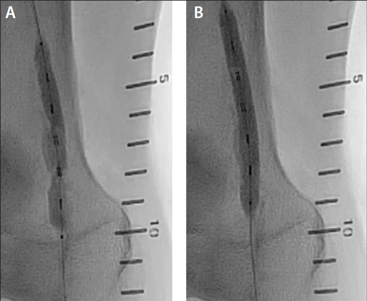
Figure 3. Highly visible emitters and fine adjustments in catheter positioning can facilitate precise calcium modification and acute lumen gain. Distal balloon marker at radiopaque number 10 in the left image (A) versus radiopaque number 9 on the right image (B).
Walk us through your clinical approach to combining SEISMIQ IVL with drug-eluting technologies such as Eluvia™ DES or Ranger™ DCB (Boston Scientific Corporation).
Dr. Golzar: It is an established clinical concept that adequate vessel preparation is a prerequisite for maximizing drug uptake. My overarching paradigm for managing femoropopliteal disease is centered on systematically preparing the vessel architecture prior to any drug delivery. The critical clinical decision tree then focuses on how you prepare the vessel, followed by the choice of antirestenotic therapy—whether a DCB or a drug-eluting stent (DES).
When using traditional atherectomy, my workflow requires a distinct, independent step: performing postatherectomy angioplasty with a standard balloon inflated to low pressures of 2 to 3 atm for approximately 60 seconds, followed by a confirmatory angiogram. IVL fundamentally streamlines this process because the system inherently combines balloon angioplasty and calcium disruption into a single step, eliminating the need for mandatory, subsequent postdilatation angioplasty.
After SEISMIQ IVL preparation, my algorithmic approach to drug delivery is guided by the post-treatment angiographic result. If there is significant residual stenosis or flow-limiting dissection, an Eluvia DES is deployed to provide structural scaffolding alongside sustained antirestenotic therapy. If there is no residual stenosis or flow-limiting dissection, a DCB is deployed and inflated at 8 atm for a full 3 minutes. This prolonged, low-to-moderate pressure inflation is critical to optimize uniform drug penetration across the fractured calcific matrix and maximize long-term vessel patency.
Dr. Stanley: My primary objectives for all lower extremity interventions are, first, to treat symptomatic hemodynamically significant lesions to achieve < 30% residual stenosis without flow-limiting dissection and, second, to deliver antiproliferative drug therapy to the treated lesions. Simply stated, these are vessel prep and drug delivery.
The primary factor driving my selection of vessel prep modality is calcium burden. In many cases, this answer is obvious, but in less severe cases without preoperative duplex or CT imaging, IVUS provides a comprehensive evaluation of the calcium present and guides my decision to use IVL. Patients with moderate-to-severe calcific plaque are treated with SEISMIQ IVL as the initial vessel prep device. Pulses are delivered throughout the calcified segments, maintaining close attention to pressure differentials during each round of therapy.
When I visualize full balloon expansion at low-pressure inflation and there is minimal pressure differential during treatment (< 0.2 atm), I move on to the next segment requiring treatment. After all stations of treatment, IVUS is repeated to assess for residual stenosis and dissection. If < 30% residual stenosis has not been achieved, additional IVL is applied to the recalcitrant lesion until the end of life of the catheter. Once < 30% residual stenosis has been achieved, I move forward with DCB angioplasty (ie, Ranger DCB) across all IVL-treated segments, utilizing a “leave nothing behind” rationale. If there is > 30% residual stenosis or flow-limiting dissection on IVUS post-IVL, selecting a DES such as Eluvia DES is a suitable option and is likely to impart a sufficient technical outcome (Figure 4 and Figure 5).
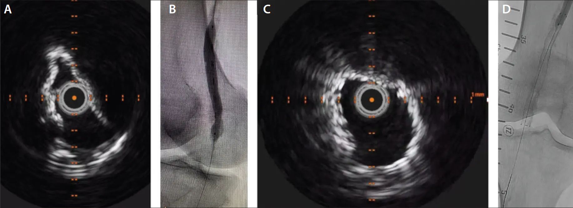
Figure 4. A diabetic patient in their early 70s with nonhealing toe wound presenting for right lower extremity intervention. Initial IVUS of a heavily calcified popliteal artery lesion (A). Treatment with SEISMIQ IVL (recalcitrant lesion noted in segment of persistent balloon stenosis) (B). IVUS of same lesion following IVL and stenting with Eluvia DES demonstrating adequate stent expansion and lumen gain (C). Fluoroscopic image of well-expanded stent in popliteal artery (D).
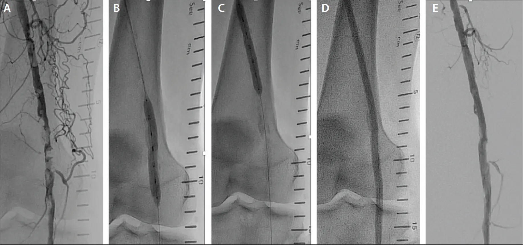
Figure 5. A man in his early 70s with Rutherford class 3 calf claudication presenting for left lower extremity angiography following a failed supervised walking program. Diagnostic angiogram demonstrating multifocal nodular severe calcific popliteal artery plaque (A). SEISMIQ IVL 6-mm catheter used at multiple stations (B, C). DCB angioplasty after IVL using Ranger 6 X 200 mm catheter (D). Completion angiogram demonstrating successful technical result (E).
Based on the randomized clinical trial designs and clinical outcomes for both DCBs and DESs, and from postmarket global registry cohorts, it is understood that drug-eluting technologies perform best when technical success (< 30% residual stenosis and no flow-limiting dissection) is realized at the index procedure.11-15 SEISMIQ IVL has demonstrated its ability to facilitate reaching these technical procedural goals even in complex and challenging cases, which gives me confidence that the chosen drug delivery method utilized in the vascular intervention is providing the ideal conditions for an optimal clinical outcome.
Furthermore, a combination of IVUS and energy delivery management with SEISMIQ IVL has so far proven to be a reliable tactic to minimize residual stenosis > 50% and poorly expanded stents, outcomes that significantly increase the potential for acute vessel thrombosis and early failure of the intervention.
In your case-based experience with SEISMIQ IVL, how has its use affected the overall resource profile of your cases?
Dr. Golzar: The 600-pulse capacity of SEISMIQ is a game-changer for cath lab economics. It allows for the comprehensive treatment of long-segment superficial femoral artery disease without the need to open additional IVL catheters, which avoids a significant increase in procedural cost. The ability to select individual treatment segments and isolate specific emitters provides a reliable mechanism to preserve pulse capacity. This targeted energy delivery ensures we can manage diffuse, multi-level calcific disease on a single device, which is a scenario that has always been a major challenge with legacy IVL technologies. Minimizing device duplication in this manner directly and favorably impacts the economic bottom line for the hospital.
Regarding the workflow enhancements, features like the intuitive touchscreen interface, zero lockout periods, and a completely sterile catheter setup that eliminates the need to bag the handle are essentially icing on the cake. By removing these clunky, manual preparation steps, the system noticeably speeds up the case, streamlines the staff’s workflow, and makes everyone’s job easier in the endovascular suite.
Dr. Stanley: Resource utilization is a constant priority in all cath labs, and as we work on more complex disease, it is important to recognize that there will be additional costs associated with the index procedure. With proper patient selection and adequate treatment principles, these additional costs can be shouldered by the system in lieu of fewer reinterventions over time. If we consider two buckets of expense for complex intervention cases—vessel prep and drug therapy—the tightrope walk for heavily calcified disease is a delicate balance between technical success and avoidance of intraprocedural complications that can send expenditures spiraling quickly out of control. Proper patient selection is crucial.
Anecdotally, I have noticed a few key differences with the use of SEISMIQ IVL in cases with severe calcium. First, I commit to front-line calcium modification with IVL without distal embolic filter as supported by trial data showing low rates of distal embolization. My reliance on atherectomy in these cases previously mandated distal filter protection, both for patient safety and cost containment. After using SEISMIQ IVL, it is becoming less common that I need to use supplementary devices for additional vessel prep. I believe this is directly related to the change in vessel wall compliance that occurs during IVL treatment. Most often, I move directly to DCB angioplasty without added steps.
Notably, at this point in the case there is an audible exhale in the lab, a diffusion of tension that is ever-present in these complex cases from the awareness of potential complications and time commitment that has been previously experienced. In a minority of scenarios where residual stenosis is suboptimal, IVUS clearly demonstrates new gaps within the calcium plaque, corresponding to adequate balloon expansion at low pressure. In these cases, a detailed analysis of the volume of residual calcium with IVUS determines the need for adjunctive treatment, such as escalation to debulking atherectomy.
There will remain a small percentage of cases that require complementary vessel prep modalities, many of which will involve IVL as one of those modalities. For these cases, we have focused on precise documentation and coding to maintain a margin that is manageable for our cath lab. Appropriate documentation to describe the complexity of the case is essential, focusing on initial diagnostic findings and, in particular, the location, severity, and extent of calcified plaque. Justification for the decision to use each treatment modality and the direct outcome after its use is equally important. Attentive coding is also of consequence, concentrating on capturing the highest level of service performed per vascular territory and using combination C-codes when applicable, which are especially suitable for IVL-inclusive procedures.
1. Brodmann M, Schlager O, Werner M, et al. Intravascular lithotripsy for peripheral artery calcification: 30-day and 6-month outcomes from the RESTORE ATK study. J Vasc Interv Radiol. 2026;37:108695. doi: 10.1016/j.jvir.2026.108695
2. Zeller T. Laser and optics-based peripheral intravascular lithotripsy system for the treatment of above-the-knee and below-the-knee calcified lesions: results of the RESTORE ATK and RESTORE BTK trials. Presented at: Vascular InterVentional Advances (VIVA); November 5, 2024; Las Vegas, Nevada.
3. Gray WA, Keirse K, Soga Y, et al. A polymer-coated, paclitaxel-eluting stent (Eluvia) versus a polymer-free, paclitaxel-coated stent (Zilver PTX) for endovascular femoropopliteal intervention (IMPERIAL): a randomised, non-inferiority trial. Lancet. 2018;392:1541-1551. doi: 10.1016/S0140-6736(18)32262-1
4. Müller-Hülsbeck S, Benko A, Soga Y, et al. Two-year efficacy and safety results from the IMPERIAL randomized study of the Eluvia polymer-coated drug-eluting stent and the Zilver PTX polymer-free drug-coated stent. Cardiovasc Intervent Radiol. 2021;44:368-375. doi: 10.1007/s00270-020-02693-1
5. Gray WA, Soga Y, Fujihara M, et al. Polymer-based drug-eluting stent treatment extends the time to reintervention for patients with symptomatic femoropopliteal artery disease: clinical evidence and potential economic value. J Comp Eff Res. 2024;13:e240025. doi: 10.57264/cer-2024-0025
6. Steiner S, Schmidt A, Zeller T, et al. COMPARE: prospective, randomized, non-inferiority trial of high- vs. low-dose paclitaxel drug-coated balloons for femoropopliteal interventions. Eur Heart J. 2020;41:2541-2552. doi: 10.1093/eurheartj/ehaa049
7. Gouëffic Y, Smith AB, Distler F, et al. Performance of stenting in femoropopliteal disease: a systematic literature review and meta-analysis of proportions. J Comp Eff Res. 2026;15:e250152. doi: 10.57264/cer-2025-0152
8. Tepe G, Brodmann M, Werner M, et al. Intravascular lithotripsy for peripheral artery calcification: 30-day outcomes from the randomized Disrupt PAD III trial. JACC Cardiovasc Interv. 2021;14:1352-1361. doi: 10.1016/j.jcin.2021.04.010
9. Tepe G, Brodmann M, Bachinsky W, et al. Intravascular lithotripsy for peripheral artery calcification: mid-term outcomes from the randomized Disrupt PAD III trial. J Soc Cardiovasc Angiogr Interv. 2022;1:100341. doi: 10.1016/j.jscai.2022.100341
10. Armstrong EJ, Adams G, Soukas PA, et al. Intravascular lithotripsy for peripheral artery calcification: 30-day outcomes from the Disrupt PAD III observational study. J Endovasc Ther. 2026;33:853-862. doi: 10.1177/15266028241283716
11. Laird JA, Schneider PA, Jaff MR, et al. Long-term clinical effectiveness of a drug-coated balloon for the treatment of femoropopliteal lesions. Circ Cardiovasc Interv. 2019;12:e007702. doi: 10.1161/CIRCINTERVENTIONS.118.007702
12. Sachar R, Soga Y, Lopez L, et al. Long-term results of the RANGER II SFA randomized trial of the Ranger drug-coated balloon vs standard percutaneous transluminal angioplasty for femoropopliteal artery treatment. Vasc Med. 2025;30:599-601. doi: 10.1177/1358863X251349782
13. Gouëffic Y, Torsello G, Zeller T, et al. Efficacy of a drug-eluting stent versus bare metal stents for symptomatic femoropopliteal peripheral artery disease: primary results of the EMINENT randomized trial. Circulation. 2022;146:1564-1576. doi: 10.1161/CIRCULATIONAHA.122.059606
14. Dake MD, Ansel GM, Jaff MR, et al. Durable clinical effectiveness with paclitaxel-eluting stents in the femoropopliteal artery: 5-year results of the Zilver PTX randomized trial. Circulation. 2016;133:1472-1483. doi: 10.1161/CIRCULATIONAHA.115.016900
15. Krishnan P, Farhan S, Schneider P, et al. Determinants of drug-coated balloon failure in patients undergoing femoropopliteal arterial intervention. J Am Coll Cardiol. 2022;80:1241-1250. doi: 10.1016/j.jacc.2022.06.043
Disclosures
Dr. Golzar: Advisory board member and speaker’s bureau for Boston Scientific Corporation.
Dr. Stanley: Advisory board member and speaker’s bureau for Boston Scientific Corporation.
Advertisement
Advertisement