Advertisement
Advertisement
July 2026
Sponsored by Penumbra, Inc.
Lightning Flash®: The Fastest Device for the Treatment of Pulmonary Embolism*
Real-world case experiences highlighting rapid clot extraction and procedural efficiency in complex pulmonary embolism and venous interventions.
With George Chrysant, MD; Matt Ramsey, MD; Tony Lu, MD; Houman Tamaddon, MD; and Brett Hyatt, DO
For decades, anticoagulation (AC) has been the cornerstone and standard of care for the treatment of acute pulmonary embolism (PE). In recent years, endovascular therapies such as thrombectomy have emerged as treatment options designed to achieve more rapid and complete thrombus removal. While thrombectomy has been prevalent in the PE space for years, there has been paucity of level 1 evidence comparing against the well-established standard of care, AC alone.
The STORM-PE randomized controlled trial has changed the conversation. As the first trial to generate level 1 evidence comparing mechanical thrombectomy to AC alone in acute intermediate-high–risk PE, STORM-PE concluded that computer assisted vacuum thrombectomy (CAVT™) plus AC demonstrated superior efficacy over AC alone.
SPEED AND EFFICACY WITHOUT COMPROMISE
The CAVT arm in STORM-PE† demonstrated a superior reduction in right heart strain compared to AC alone, with a mean change in right ventricular/left ventricular (RV/LV) ratio of 29.7% versus 13.1% at 48 hours (P < .001).1 Safety was comparable between groups with composite major adverse event (MAE) rates being 4.3% in the CAVT arm versus 7.5% in the AC arm, with zero device-related transfusions in the CAVT arm.1‡
CAVT device performance was also reflective of how technically evolved thrombectomy devices have become. The median device time for the CAVT arm was 25 minutes—the fastest of any PE-indicated mechanical thrombectomy device (Figure 1)—with a median total procedure time of 56 minutes. Technical success was achieved in 100% of cases.1
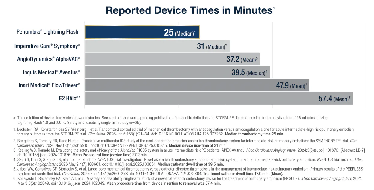
Figure 1.
PATIENTS EXPERIENCE FUNCTIONAL RECOVERY FASTER WITH CAVT
The 90-day STORM-PE data, presented at SIR 2026, include functional recovery and quality-of-life outcomes. CAVT patients demonstrated 12 times higher odds of achieving New York Heart Association (NYHA) class I status (no physical limitations) compared to AC alone.2 The 6-minute walk test showed CAVT patients walking 472 m versus 376 m with AC at 90 days—a statistically significant difference of 96 m, roughly the length of a football field.3 For patients, these gains translate into tangible daily activities, such as exercise, running errands, and returning to pre-PE status.
LIGHTNING FLASH 3.0: IMPROVEMENTS IN DESIGN, DETECTION, AND SPEED
From a speed perspective, Lightning Flash 3.0 shows a clear step forward with 1.3 times faster clot removal.§ The device’s larger lumen tubing is engineered to reduce systematic friction from ingested thrombus, maintaining full vacuum power at the catheter tip. An automated decompression feature further mitigates friction buildup within the tubing, minimizing thrombus-related obstructions during aspiration, allowing for shorter procedure times.
Lightning Flash 3.0 is designed to mitigate blood loss frontline, streamlining procedural workflow by eliminating the need for blood return strategies.
This improvement stems from enhanced algorithmic sensitivity and a notable hardware change: relocating the clot detection computer from the top of the canister to a position closer to the CAT16 aspiration catheter (Penumbra, Inc.; Figure 2). Positioning the computer closer to the point of thrombectomy enables the system to rapidly distinguish thrombus from patent flow. The result is 60% fluid savings§ without compromising efficacy.
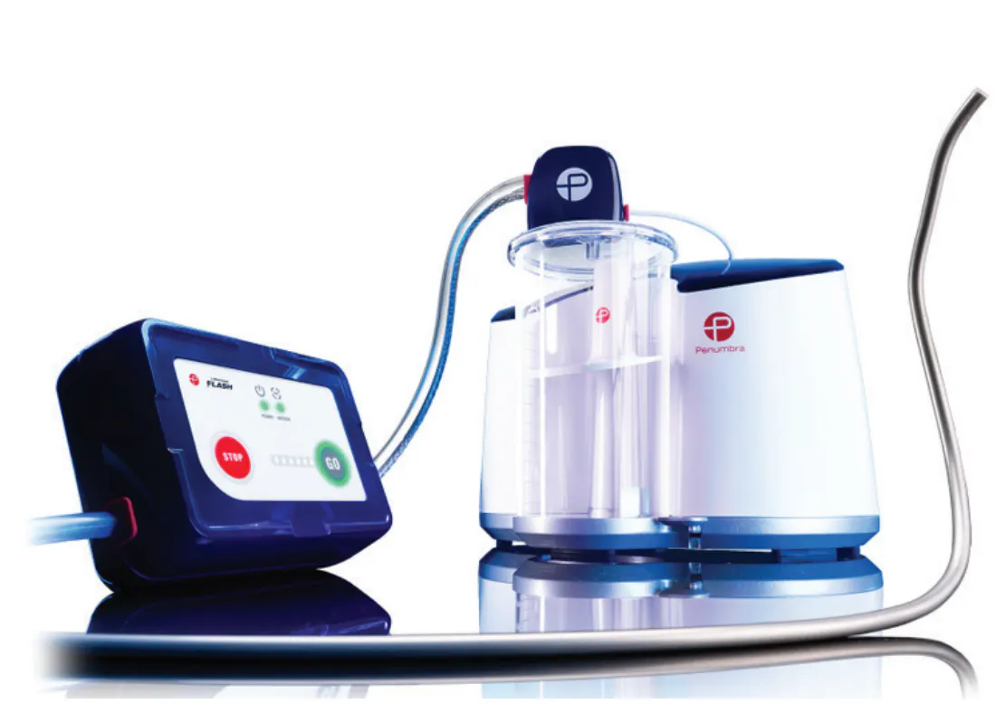
Figure 2. Lightning Flash 3.0 and CAT16 aspiration catheter.
Workflow simplicity rounds out the system with the updated Lightning Flash console, providing clear, streamlined audiovisual feedback with an intuitive layout. Integrated air detection and straightforward operation reduce technologic complexity, allowing clinicians to focus where it matters most: on the patient and the procedure.
Lightning Flash, coupled with the Element™ Vascular Access System (Penumbra, Inc.), is designed to enhance procedural efficiency. Penumbra’s 17-F Element Vascular Access System is engineered to provide seamless and stable access to the pulmonary and venous vasculature. Together, Lightning Flash and Element form a cohesive thrombectomy platform designed to facilitate efficient removal of large thrombus.
*See Figure 1.
†The devices included in STORM-PE are Lightning Flash 1.0 and Lightning Flash 2.0.
‡STORM-PE was powered for the primary endpoint only.
§Compared to Lightning Flash 2.0. Tests performed and data on file at Penumbra, Inc. Test performed using bovine blood and water. Bovine blood took 1.3x more time to be fully ingested in bench top testing of Lightning Flash 2.0 when compared to Lightning Flash 3.0, while 60% less water was removed with Lightning Flash 3.0 when compared to Lightning Flash 2.0. Bench test results may not be indicative of clinical performance.
1. Lookstein RA, Konstantinides SV, Weinberg I, et al. Randomized controlled trial of mechanical thrombectomy with anticoagulation versus anticoagulation alone for acute intermediate-high risk pulmonary embolism: primary outcomes from the STORM-PE trial. Circulation. 2026;153:21-34. doi: 10.1161/CIRCULATIONAHA.125.077232
2. Lookstein R. Clinical, functional, and quality of life outcomes through 90 days in the STORM-PE RCT for mechanical thrombectomy with anticoagulation vs anticoagulation alone in acute intermediate-high risk PE patients. Presented at: Society of Interventional Radiology Annual Scientific Meeting (SIR); April 11-15, 2026; Toronto, Canada.
3. Rosovsky RP. Randomized controlled trial of mechanical thrombectomy with anticoagulation versus anticoagulation alone for acute intermediate-high risk PE: primary outcome, functional endpoints, and core lab findings from STORM-PE. Presented at: Vascular InterVentional Advances Conference (VIVA) November 2-5, 2025; Las Vegas, NV.
High-Risk PE Management With Lightning Flash 3.0

Patient Presentation
A man in his early 70s presented with acute PE symptoms consistent with American College of Cardiology/American Heart Association (ACC/AHA) class D2: incipient cardiopulmonary failure with normotensive shock, confirmed by CT imaging and positive biomarkers. This required escalation of care and transfer to a tertiary center. The patient developed respiratory compromise requiring high-flow bilevel positive airway pressure and was initiated on extracorporeal membrane oxygenation (ECMO) support. Treatment decisions included emergent CAVT using the Lightning Flash 3.0 system while on ECMO.
Intervention
After gaining access through femoral vein, an Element vascular access sheath was placed. Pulmonary angiography revealed extensive bilateral PE with a large clot burden involving the right pulmonary artery (PA; Figure 1), including upper, middle, and lower lobes, as well as the lower lobe branches of the left PA (Figure 2).
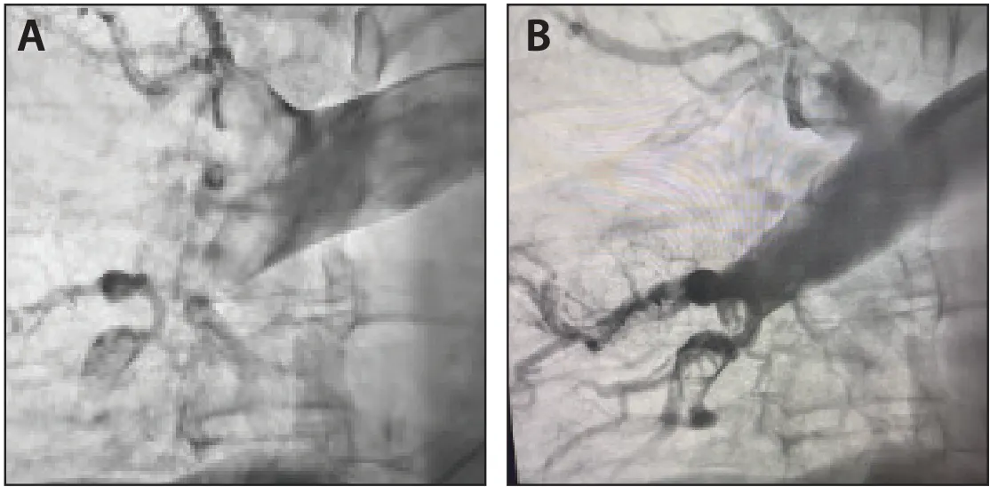
Figure 1. Pre- (A) and post-thrombectomy (B) right PA angiogram.
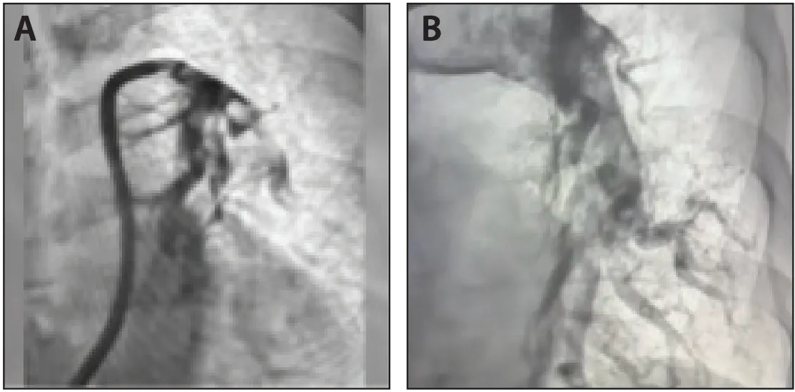
Figure 2. Pre- (A) and post-thrombectomy (B) left PA angiogram.
The CAT16 aspiration catheter was advanced, and thrombectomy was performed. Rapid and effective aspiration resulted in removal of a massive clot burden from the right and left PA territories. Immediate improvement in perfusion was observed with restoration of flow and angiographic blush across treated pulmonary segments. Total device time was 7 minutes and 15 seconds, and total case time was 44 minutes.
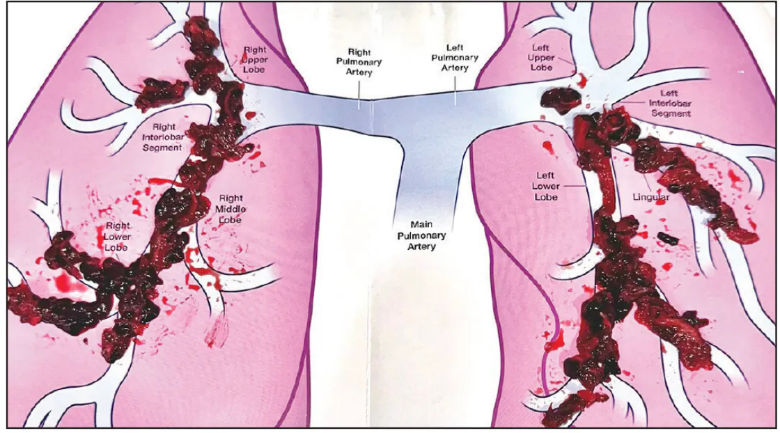
Figure 3. Thrombus aspirated.
Discussion
This case demonstrates successful use of CAVT in a critically ill patient with massive PE requiring ECMO support. The patient experienced immediate hemodynamic and perfusion improvement following clot removal, and was weaned to room air and decannulated from ECMO within 24 hours. The Lightning Flash 3.0 system, supported by the Element sheath, enabled rapid and effective thrombus removal, highlighting its utility in high-acuity PE cases and supporting its relevance for advanced cases.
Bilateral Saddle PE Treatment With Lightning Flash 3.0

Note: This case was performed at Duke University Hospital in Durham, North Carolina.
Patient Presentation
The patient presented with shortness of breath and tachycardia but remained hemodynamically stable. CT and angiographic imaging demonstrated a high-clot-burden, bilateral PE with a large saddle component. Pulmonary thrombectomy was performed using Penumbra’s CAVT with the Lightning Flash 3.0 system.
Intervention
The PAs were accessed using a pigtail catheter and J-wire, which was then exchanged for an 1-cm flexible tip, 260-cm-long Amplatz Super Stiff™ guidewire (Boston Scientific Corporation). With the Amplatz wire in place in the right PA, the team exchanged the initial sheath for the 65-cm Element vascular access system. The Element sheath’s hypotube composition facilitated access through the right heart and past the pulmonic valve until positioned into the main PA (Figure 1).
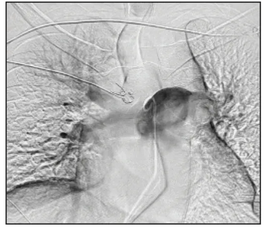
Figure 1. Pre-pulmonary angiogram.
With the Element sheath in place, the CAT16 aspiration catheter was advanced into the PAs, and aspiration thrombectomy was performed. Clot removal was carried out systematically from proximal to distal segments, beginning centrally in the right segment followed by the lower right segments. Once the right side was effectively treated, the aspiration catheter was transitioned over a wire to the left PAs for treatment of the left main PA and lobar branches. After completion of thrombectomy, a substantial volume of thrombus was removed (Figure 2 and Figure 3).
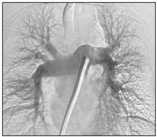
Figure 2. Post-thrombectomy pulmonary angiogram.
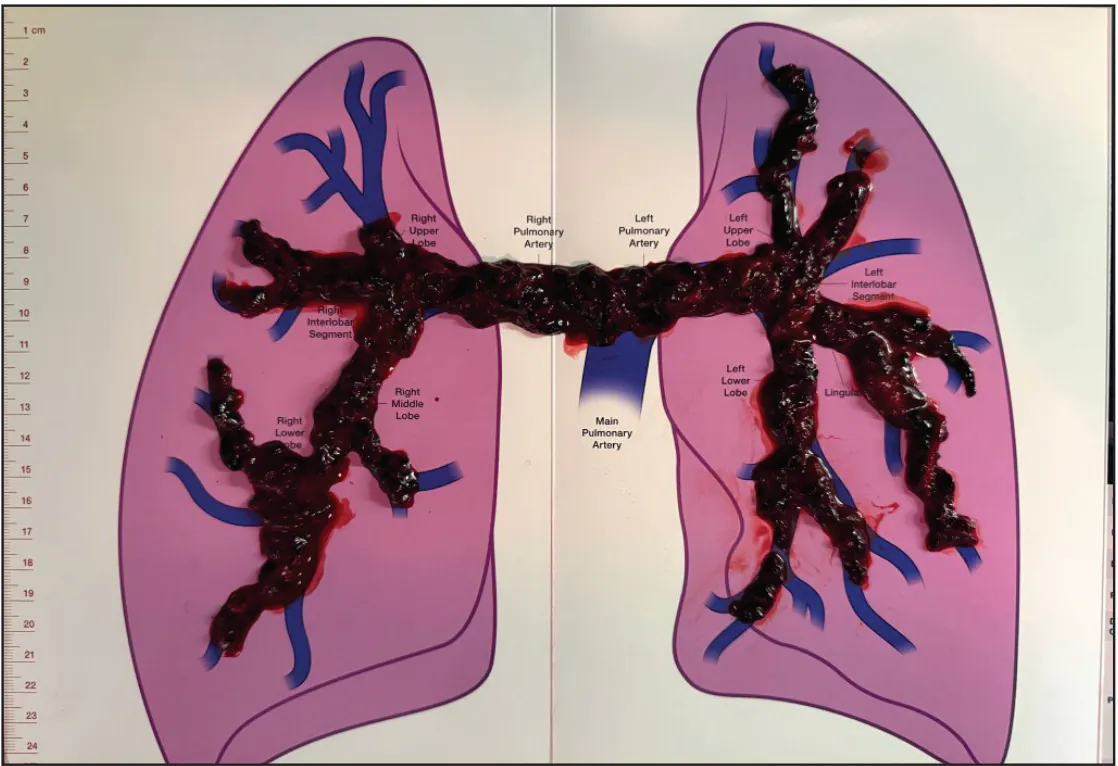
Figure 3. Thrombus aspirated.
Discussion
The patient experienced a significant hemodynamic improvement, with mean PA pressures (mPAPs) decreasing from 31 mm Hg preprocedure to 20 mm Hg postprocedure. The Lightning Flash system, supported by the Element sheath, enabled efficient removal of extensive bilateral clot burden with restoration of pulmonary perfusion and rapid clinical improvement. Despite removing a significant amount of thrombus, the system’s clot-detection algorithm mitigated the risk of high estimated blood loss frontline, by accurately transitioning from full aspiration mode when embedded in thrombus and sampling mode when in patent flow.
Bilateral PE Management With Penumbra’s Complete PE Platform

Patient Presentation
A woman in her early 70s presented with 5 days of worsening dyspnea at rest and a 4 L/min oxygen requirement. Her history included hypertension, dyslipidemia, chronic kidney disease, morbid obesity, as well as a prior PE (previously treated with catheter-directed thrombolytic therapy). The patient also reported a 1-week history of right knee pain with active and passive motion and was found to have an ipsilateral, nonocclusive popliteal deep vein thrombosis. CT and pulmonary angiography demonstrated extensive central, lobar, and segmental PE (Figure 1A and Figure 2A) with a RV/LV ratio of 3.0. Additionally, transthoracic echocardiogram revealed severely reduced systolic function. The patient was started on therapeutic AC, and a decision was made to proceed with aspiration thrombectomy using the Penumbra Lightning Flash 3.0 System.
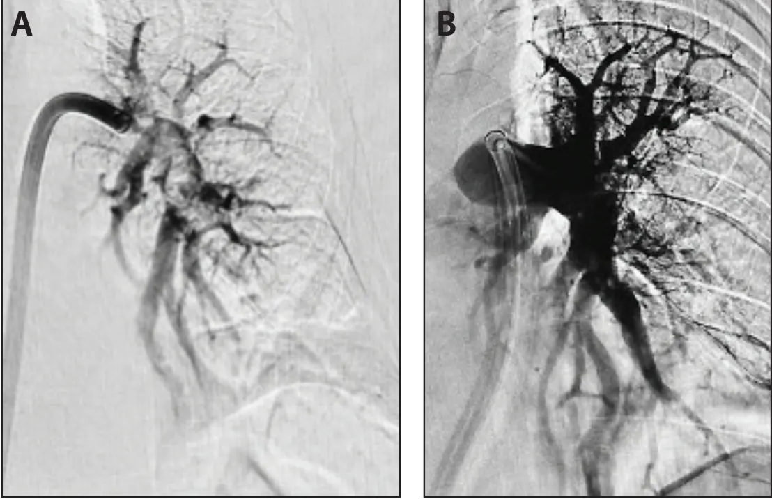
Figure 1. Pre left angiogram (A). Post-thrombectomy angiogram (B).
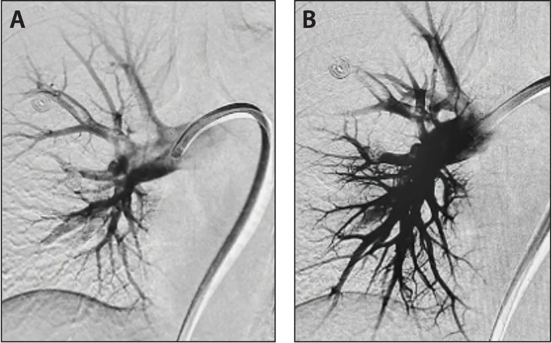
Figure 2. Pre right angiogram (A). Post-thrombectomy angiogram (B).
Intervention
The right common femoral vein was accessed under ultrasound guidance, and an angled pigtail catheter was used to cross the right heart and access the main PA. The right and left PAs were then selectively catheterized using a KMP catheter and Glidewire® (Terumo® Interventional Systems), which were exchanged for a short-tip Amplatz stiff guidewire to facilitate advancement of a 65-cm Element sheath. Starting PA pressures were found to be 57/27 mm Hg (mPAP, 37 mm Hg). Aspiration thrombectomy was performed using the Lightning Flash 3.0 System until satisfactory reduction in thrombus burden and improvement in hemodynamics were achieved (Figure 1B and Figure 2B). The total procedure time was 63 minutes with an estimated blood loss of 200 mL.
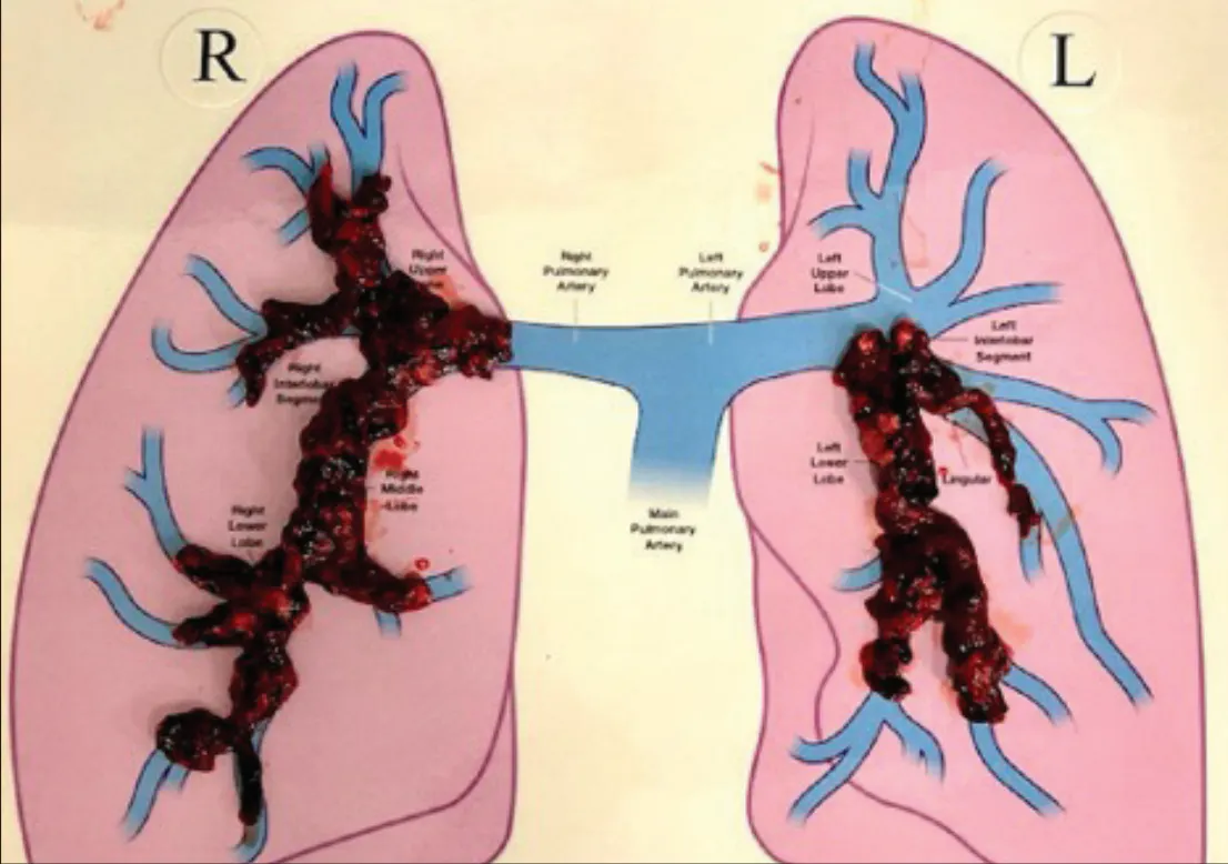
Figure 3. Thrombus aspirated.
Discussion
The patient’s final PA pressures were 41/20 mm Hg (mPAP, 28 mm Hg), a mPAP decrease of 9 mm Hg while still on the table. The patient was downgraded to floor status and weaned off supplemental oxygen overnight. On postprocedure day 1, she was able to ambulate 150 ft before fatiguing and was transitioned to lifelong oral AC. Lightning Flash 3.0 enabled the patient to achieve significant clinical improvement within 24 hours and proved to be a valuable tool in the treatment of PE.
High-Volume Bilateral Venous Thrombus Removal From the IJ Approach

Patient Presentation
An adult patient with a history of PE and indwelling inferior vena cava (IVC) filter presented with bilateral lower extremity swelling and pain due to lower extremity venous thrombus, along with IVC filter malfunction and IVC thrombosis. Management decisions included catheter-directed thrombectomy of the bilateral lower extremities, IVC removal of the malfunctioning filter, and placement of a new IVC filter. Considering the suspected extensive nature of the thrombus as well as both popliteal veins being occluded, the decision was made to access via the internal jugular (IJ) vein so that the iliofemoral, popliteal and inflow vessels, such as the profunda and saphenous, of both legs could be accessed.
Intervention
After gaining access via the right IJ vein under ultrasound guidance, existing central venous access was rewired and upsized to an 8-F sheath. Venography demonstrated extensive thrombosis of the IVC, iliac, femoral, popliteal, and great saphenous veins bilaterally, with thrombus extending around and above the IVC filter. A 17-F Element sheath was advanced into the IVC, and the Lightning Flash 3.0 system was utilized for aspiration thrombectomy.
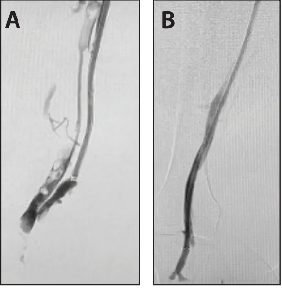
Figure 1. Pre- (A) and postthrombectomy (B) angiogram.
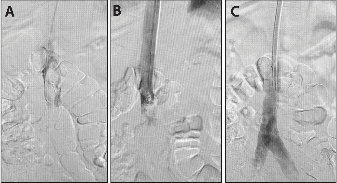
Figure 2. Pre- (A), intra- (B), and post- (C) thrombectomy cavagram.
Initial thrombectomy was performed in the suprarenal and infrarenal IVC to reduce clot burden and mitigate embolic risk. The device was then sequentially advanced into the right iliac, femoral, profunda, popliteal, and great saphenous veins, achieving substantial thrombus removal and restoration of flow. The left-sided venous system was managed similarly.
Due to persistent occlusion within the existing IVC filter, the filter was removed using a retrieval system. Further thrombectomy of the IVC was performed, followed by placement of a new Celect Platinum® IVC filter (Cook® Medical) in the perirenal position. Completion venography demonstrated improved flow through targeted segments.
Discussion
Successful aspiration thrombectomy resulted in significant reduction of thrombus burden across the IVC and bilateral lower extremity venous systems (Figure 3), with restoration of venous patency in most segments. The malfunctioning IVC filter was safely removed and replaced. The flexibility, aspiration power, and trackability of the Lightning Flash 3.0 system, combined with the support of the Element sheath, enabled effective management of extensive venous thrombosis with favorable angiographic outcomes.
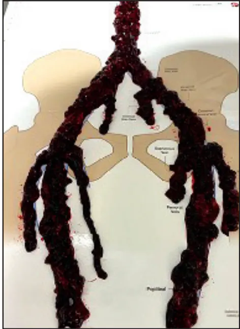
Figure 3. Thrombus aspirated.
Unilateral Venous Thrombectomy With Caval Involvement With Lightning Flash 3.0

Patient Presentation
The patient presented to the hospital with lower extremity swelling and pain. Initial diagnostic venous compression ultrasound confirmed the suspected presence of occlusive thrombus in the left leg. Considering how symptomatic the patient was, the decision was made to proceed with thrombectomy for immediate thrombus resolution.
Intervention
After gaining access in the left popliteal vein under ultrasound guidance, the team began initial imaging. Venography confirmed an extensive occlusion running from the popliteal vein through the iliofemoral and into the IVC where a previously placed IVC filter was noted. The level of collaterals present upon contrast injection suggested a high probability that the thrombus was composed of a more organized morphology (Figure 1A and 1C).
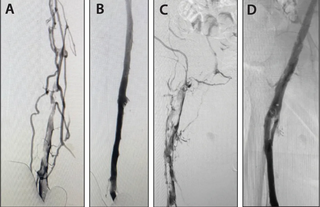
Figure 1. Pre- (A) and postprocedural (B) femoral intervention venogram. Pre- (C) and postprocedural (D) iliac intervention venogram.
With the target segments identified through venography, the Lightning Flash 3.0 aspiration catheter was advanced into the vasculature. Upon introduction into the popliteal vein, aspiration was initiated almost immediately. The aspiration catheter was advanced antegrade in a proximal-to-distal fashion. Upon evacuation of the thrombus in the popliteal to iliac vein, focus was shifted to the caval thrombus. The target thrombus was removed within a few seconds of aspiration in the IVC, with only the more stubborn clot remaining in the filter. The 1:1 torqueability of the CAT16 aspiration catheter was advantageous in allowing the tip of the catheter to sweep effectively through the filter, enabling complete thrombus resolution prior to removal (Figure 1B and 1D and Figure 2).
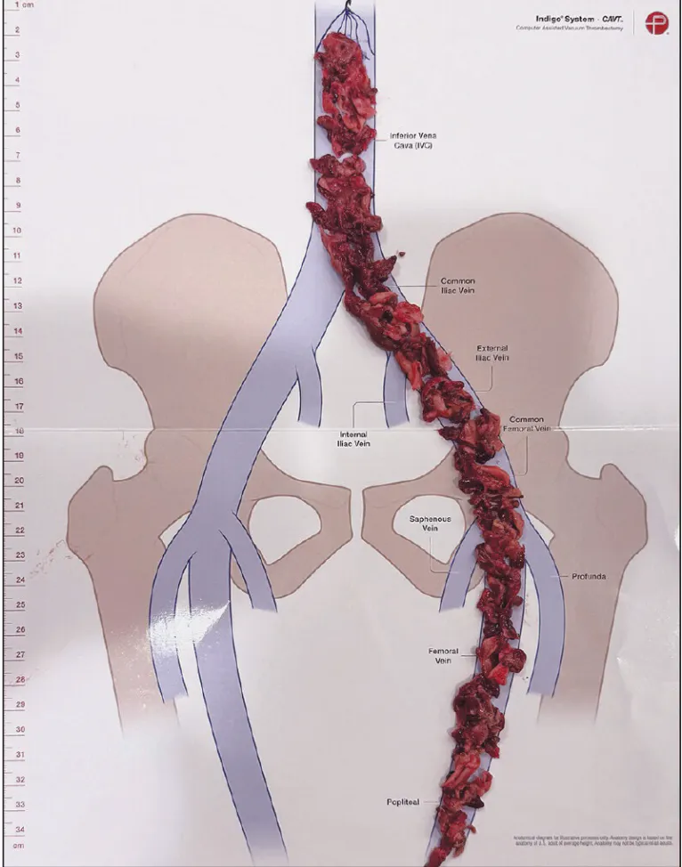
Figure 2. Thrombus aspirated.
Discussion
The Lightning Flash 3.0 system successfully reduced extensive thrombus burden from the popliteal to caval segments, restoring venous flow. Lightning Flash 3.0 enabled effective clot removal and revascularization in a complex, multi-segment venous occlusion.
Disclaimer: The opinions and clinical experiences presented herein are for informational purposes only. The results may not be predictive of all patients. Individual results may vary depending on a variety of patient-specific attributes.
Advertisement
Advertisement