Advertisement
Advertisement
July 2026
Sponsored by Medtronic
One Size Does Not Fit All: Beyond Anatomy in Pelvic Venous Disease

Pelvic venous disease (PeVD) is estimated to occur in up to one-third of women and can significantly impact quality of life (QOL).1,2 Patients often present with venous-origin chronic pelvic pain (VO-CPP), dyspareunia, dysmenorrhea, lower limb venous incompetence, or extrapelvic varices, and they undergo a multitude of office visits before obtaining symptom relief.1 PeVD with VO-CPP is underrepresented in global prevalence studies, further highlighting the need for increased awareness and comprehensive, individualized routine care.2,3 Although 2023 guidelines published by the Society for Vascular Surgery, American Venous Forum, and American Vein and Lymphatic Society acknowledged the efficacy of gonadal/ovarian vein embolization (GVE) for symptom relief in patients with pelvic varicosities, more research is needed in this disease space.4
Dr. Knuttinen, an expert in the field of vascular radiology, shares her experience and perspective on complex PeVD diagnostics and individualized, longitudinal treatment.
CASE STUDY: TO EMBOLIZE OR STENT?
A patient was referred to our practice for GVE based on cross-sectional imaging findings of primary pelvic venous insufficiency. The treatment pathway seemed straightforward. During diagnostic venography and contrast, the renal and gonadal veins demonstrated preserved valvular competency without significant reflux (Figure 1A). However, further evaluation demonstrated hemodynamically significant left common iliac vein compression with extensive pelvic collaterals (Figure 1B). Rather than GVE, iliac vein stenting with the Abre™ venous self-expanding stent system (Medtronic) was performed, resulting in symptom improvement. What initially appeared to be a routine GVE referral ultimately altered our treatment strategy completely.
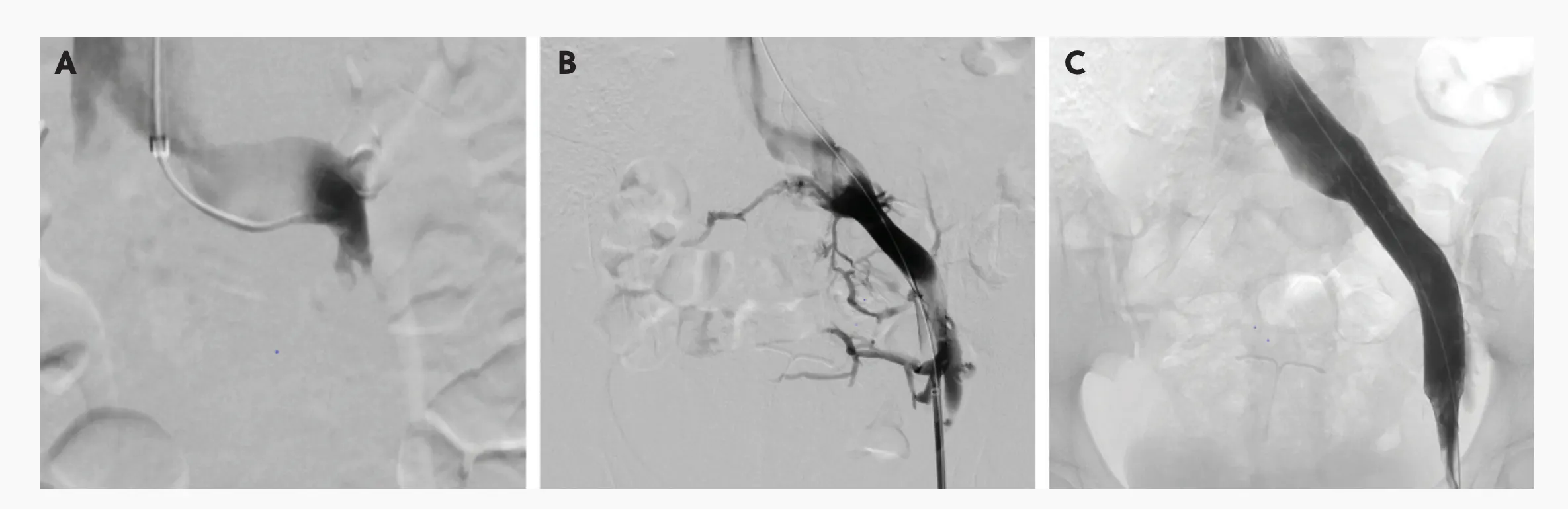
Figure 1. Venography before and after iliac vein stenting. Initial venography showed preserved valvular competency of the left renal vein (A). The pelvic collaterals resulting from the iliac vein compression (B). Restoration of flow in the left common iliac vein poststenting (C).
APPROPRIATE PeVD DIAGNOSIS AND CASE DISCUSSION
Diagnostic venography with intravascular ultrasound (IVUS) provides information that cannot be fully appreciated on cross-sectional imaging alone. Importantly, not all gonadal venous reflux represents a primary embolization target. In selected patients, gonadal venous insufficiency may function as a decompressive collateral pathway secondary to an underlying deep venous obstruction. Heightened central sensitization and altered pain processing through elevated neuropeptides may potentially help explain why symptom severity often correlates poorly with imaging findings and why treatment responses vary among individuals.5 These observations have reinforced the importance of longitudinal patient engagement, expectation setting, and multidisciplinary care.
Imaging studies don’t tell the whole picture. The patient described earlier serves as an important example of this. What initially appeared to be a straightforward embolization ultimately proved to be a manifestation of significant iliac venous outflow obstruction.
TREATMENT CONSIDERATIONS
We have found that individualized consultation and direct patient engagement are critical in establishing an appropriate treatment strategy (Figure 2). Direct review of imaging with patients often improves shared decision-making and allows patients to better understand the basis of their symptoms.
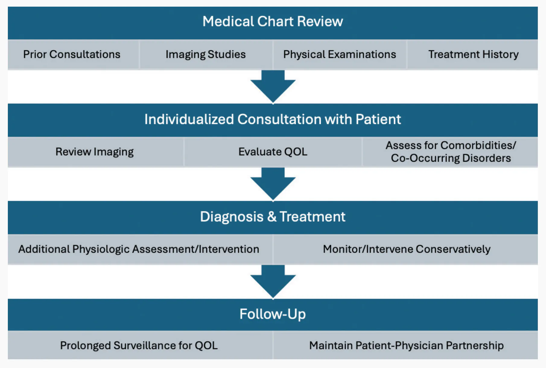
Figure 2. Our treatment strategy has developed over the years to capture a more individualized and holistic approach.
Importantly, not every patient with pelvic venous reflux requires intervention. Patients with minimal QOL impairment may be managed conservatively, whereas patients with substantial QOL limitations may benefit from further physiologic assessment to determine whether embolization, endovascular stenting, or combined approaches should be considered. Cases such as this one emphasize the importance of physiologic evaluation and individualized treatment planning in patients with VO-CPP. Although GVE is an important and successful treatment strategy, cross-sectional imaging may not fully characterize venous flow dynamics or collateralization patterns. Careful interpretation of venography and IVUS findings can help guide appropriate intervention while avoiding reliance on a uniform treatment algorithm.
This concept aligns with emerging literature demonstrating symptomatic improvement following primary iliac venous stenting in carefully selected patients.6 Close longitudinal follow-up remains essential to evaluate venous hemodynamics and, more importantly, to assess improvement in patient symptoms and QOL.
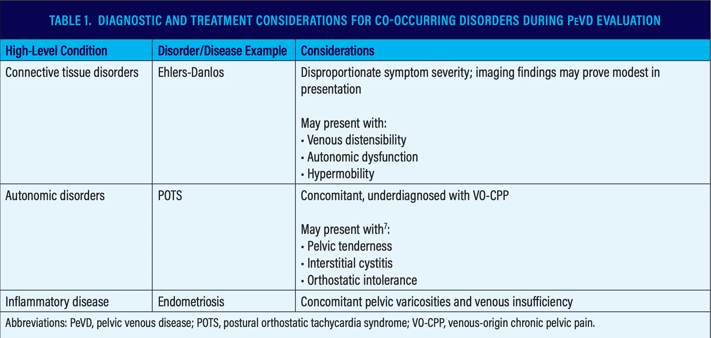
In younger patients and patients with hypermobility spectrum disorders, the development of patient-physician partnership becomes increasingly important given the potential need for prolonged surveillance after intervention (Table 1).7 In these patients, careful consideration of stent sizing, venous elasticity, and long-term crush resistance is critical when planning treatment strategies.
REFLECTIONS
Earlier in our experience, treatment decisions were often driven by anatomic findings, such as dilated gonadal veins or enlarged pelvic varices. However, over time we have come to appreciate that physiology matters just as much. Venous hemodynamics, pain processing, autonomic dysfunction, connective tissue disorders, and patient-specific factors can all influence symptom development and treatment response.
In our experience, the most symptomatic patients are not always those with the most dramatic imaging findings. This disconnect has encouraged us to move away from strict treatment algorithms and toward a more holistic individualized approach to patient evaluation (Figure 2).
KEY TAKEAWAYS
Ultimately, the management of PeVD requires recognition that VO-CPP is not a “one-size-fits-all” condition. The future of PeVD management will likely depend less on standardized treatment algorithms and more on individualized physiologic interpretation, multidisciplinary collaboration, and meaningful partnerships with patients. As our understanding continues to evolve, perhaps the most important lesson remains the simplest: Every patient’s story is different.
1. Zondervan KT, Yudkin PL, Vessey MP, et al. Prevalence and incidence of chronic pelvic pain in primary care: evidence from a national general practice database. Br J Obstet Gynaecol. 1999;106:1149-55. doi: 10.1111/j.1471-0528.1999.tb08140.x
2. Ahangari A. Prevalence of chronic pelvic pain among women: an updated review. Pain Physician. 2014;17:E141-7.
3. Kavallieros K, Pope T, Tan M, et al. Identification of outcomes in clinical studies for pelvic venous disorders. J Vasc Surg Venous Lymphat Disord. 2024;12:101865. doi: 10.1016/j.jvsv.2024.101865
4. Gloviczki P, Lawrence PF, Wasan SM, et al. The 2023 Society for Vascular Surgery, American Venous Forum, and American Vein and Lymphatic Society clinical practice guidelines for the management of varicose veins of the lower extremities. Part II: endorsed by the Society of Interventional Radiology and the Society for Vascular Medicine. J Vasc Surg Venous Lymphat Disord. 2024;12:101670. Published correction appears in J Vasc Surg Venous Lymphat Disord. 2024;12:101923. doi: 10.1016/j.jvsv.2023.08.011
5. Gavrilov SG, Vassilieva GY, Vasilev IM, Grishenkova AS. The role of vasoactive neuropeptides in the genesis of venous pelvic pain: a review. Phlebology. 2020;35:4-9. doi: 10.1177/0268355519855598
6. Murali N, Gupta R, Desai KR. The role of iliac vein stent placement in pelvic venous disorder management. J Vasc Surg Venous Lymphat Disord. 2024;12:101696. doi: 10.1016/j.jvsv.2023.101696
7. Smith SJ, Sichlau MJ, Smith BH, et al. Improvement in chronic pelvic pain, orthostatic intolerance and interstitial cystitis symptoms after treatment of pelvic vein insufficiency. Phlebology. 2024;39:202-213. doi: 10.1177/02683555231219737
Disclosures
Dr. Knuttinen: Consultant to Innova, Medtronic, and Penumbra.
Abre™ venous self-expanding stent system Brief Statement
Intended Use/Indications: The Abre™ venous self-expanding stent system (Abre™ stent system) is indicated for use in the iliofemoral veins for the treatment of symptomatic venous outflow obstruction.
Contraindications: Do not use the Abre™ stent system with patients with known hypersensitivity to nickel titanium (nitinol), with patients who are judged to have a lesion that prevents complete inflation of a balloon dilatation catheter or proper placement of the stent or the stent delivery system, and with patients in whom anticoagulant or antiplatelet therapy is contraindicated.
Potential Adverse Effects of the Device on Health: The potential adverse effects (e.g., complications) associated with the use of the Abre™ stent system include, but are not limited to, access failure, access site infection, allergic reaction to contrast medium or procedure medications; aneurysm; AV fistula; bleeding; brucourtesyng; death; device breakage; device maldeployment; edema; embolization; fever; hematoma; hypertension; hypotension, nausea, or other vasovagal response; infection; myocardial infarction, arrhythmia, or other cardiovascular insufficiency; open surgical repair; pain; pseudoaneurysm; renal insufficiency or renal failure (new or worsening); respiratory distress or pulmonary embolism; sepsis; stent fracture; stent malapposition; stent malposition; stent migration; stroke, paradoxical embolism, transient ischemic attack, or intracerebral hemorrhage; tissue necrosis; venous occlusion, restenosis, or thrombosis, within or outside of stented segment; and vessel damage, including intimal injury, dissection, perforation, or rupture.
Warnings, precautions, and instructions for use can be found in the product labeling at http://manuals.medtronic.com.
CAUTION: Federal (USA) law restricts this device to sale by or on the order of a physician.
US-CV-2601575 ©2026 Medtronic. Medtronic, Medtronic logo are trademarks of Medtronic. All other brands are trademarks of Medtronic. For global distribution. 07/2026
Advertisement
Advertisement