Advertisement
Advertisement
August 2026
Sponsored by Johnson & Johnson Med Tech
Shockwave IVL’s Pioneering Mechanism of Action: Unique Acoustic Waveform, Consistent Output
A closer look at Shockwave™ IVL’s unique acoustic waveform and its circumferential, longitudinal, and pulse-to-pulse consistency.
Intravascular lithotripsy (IVL) is the energy-based production of ultrasonic acoustic pressure waves for the modification, fracture, and fragmentation of vascular calcification. Since its commercial introduction in 2017, Shockwave IVL (Shockwave Medical, now a part of J&J Med Tech) has become the preferred treatment strategy among physicians treating calcific peripheral artery disease. An underappreciated aspect of Shockwave IVL is its mechanism of action, which consists of a tuned acoustic waveform and a consistent ultrasonic acoustic output that mechanistically explain Shockwave IVL’s clinically validated predictable safety and consistent efficacy.1 This mechanistic foundation is likely to become a critical point of differentiation as new IVL technologies come to market with similar-looking platforms.
SHOCKWAVE’S ACOUSTIC WAVEFORM: TUNED TO PRIORITIZE SAFETY WITHOUT TRADEOFFS FOR EFFICACY
Prior to the commercialization of its first device, Shockwave Medical spent nearly a decade tuning ultrasonic acoustic pressure waves, originally used for kidney stone management, for use in the arterial space. Shockwave IVL modifies calcium through the energy-based production of Shockwaves, which are a specialized form of ultrasonic acoustic pressure waves tuned to prioritize safety without tradeoffs for efficacy. Shockwave’s acoustic pressure waveform consists of a very fast transition and high-amplitude positive peak pressure, which is responsible for creating compressive, shear, squeezing, and other forces that modify calcium, followed by a low-amplitude, negative peak pressure that minimizes soft tissue damage from tensile stress (Figure 1).2 Shockwave IVL’s unique and published acoustic waveform is a key component to its robust and growing body of clinical evidence of > 25,000 published patient outcomes across > 600 journals, demonstrating predictable safety and consistent efficacy.3
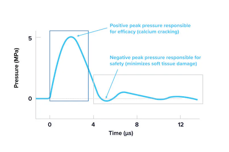
Figure 1. Shockwave IVL’s unique acoustic pressure waveform is tuned to prioritize safety without tradeoffs for efficacy across all products.
SHOCKWAVE IVL’S CIRCUMFERENTIAL ENERGY DELIVERY
Shockwave IVL’s catheter design consists of a series of emitters with two 180° spaced spark gaps per emitter. Energy travels from the generator and is delivered at the spark gaps, launching two initial Shockwaves (one per spark gap) that merge to create a spherical pressure field around each emitter. The ultrasonic acoustic output of the spherical Shockwave produced is distributed circumferentially around the balloon edge, lacking dead zones or areas with an acoustic output less than the threshold to modify calcium (Figure 2).4 Shockwave IVL’s emitter design prioritizes device delivery and crossing performance while maintaining clinical efficacy across all types of calcium.5,6
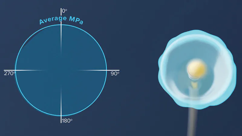
Figure 2. The ultrasonic acoustic output of the spherical Shockwave field is distributed circumferentially around the balloon edge.
SHOCKWAVE IVL’S LONGITUDINAL ACOUSTIC OUTPUT AND PULSE-TO-PULSE CONSISTENCY
Shockwave IVL devices consist of strategically placed, tandem-firing emitters that ensure a consistent acoustic output along the length of the device to disrupt calcium regardless of where it is located along the catheter. While the area of highest acoustic output is adjacent to the emitters, the acoustic output of Shockwave E8, Shockwave’s 80-mm device for calcific disease above and below the knee, is measured to be therapeutic along the balloon edge and clinically relevant up to 6 mm away from the emitter, allowing for the modification of superficial, deep, and medial calcification (Figure 3).7
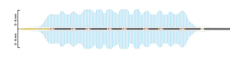
Figure 3. Shockwave E8’s acoustic output is therapeutic along the balloon edge and clinically relevant up to 6 mm away from emitters.
One pulse from a Shockwave IVL device activates two emitters in tandem, producing two spherical Shockwaves per pulse. The benefits of Shockwave IVL’s tandem-firing emitters are twofold. First, the simultaneously produced Shockwaves intersect between the tandem-firing emitters at the balloon edge, resulting in shock-shock interaction. Shock-shock interaction facilitates a more constant acoustic pressure across the balloon edge as compared to independent-firing emitters.8 Secondly, tandem-firing emitters maximize treatment capability. Because two Shockwaves are created with each pulse, Shockwave E8’s 400 available pulses per catheter provide physicians 800 Shockwaves for modification fracture and fragmentation of calcification. Moreover, these 800 Shockwaves provide a consistent acoustic output from the initial to last Shockwaves available with 98% to 102% WavePower consistency (Figure 4).9,10
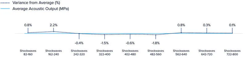
Figure 4. The acoustic output of the 800 available Shockwaves from Shockwave E8 are consistent from the initial to last Shockwaves available.
WHY THIS MATTERS FOR THE FUTURE STATE OF IVL
Shockwave IVL’s clinically validated safety and consistent efficacy are rooted in the fundamentals of its mechanism of action: a tuned acoustic waveform designed to modify calcium while protecting soft tissue, circumferential energy delivery without dead zones, longitudinal treatment coverage along the catheter, and pulse-to-pulse consistency from the initial to last Shockwaves available per catheter. Together, these features help explain Shockwave IVL’s reliable performance and consistent efficacy within real-world patient populations and complex lesion types.11 Finally, Shockwave IVL is committed to continuing to earn its innovative disruptor reputation by building upon its clinically validated mechanistic foundation and advancing future IVL solutions across new indications and disease states to benefit physicians and, ultimately, the patients they treat.
1. Tepe G, Brodmann M, Werner M, et al. Intravascular lithotripsy for peripheral artery calcification: 30-day outcomes from the randomized Disrupt PAD III trial. JACC Cardiovasc Interv. 2021;14:1352-1361. doi: 10.1016/j.jcin.2021.04.010
2. Kereiakes DJ, Virmani R, Hokama JY, et al. Principles of intravascular lithotripsy for calcific plaque modification. JACC Cardiovasc Interv. 2021;14:1275-1292. doi: 10.1016/j.jcin.2021.03.036
3. Bibliography available upon request.
4. Data on file, Shockwave Medical. Based on by quadrant average acoustic output of 28 measurements at balloon edge adjacent to emitter. IEC standards 62127 & 61846.
5. Ali ZA, Kereiakes DJ, Hill JM, et al. Impact of calcium eccentricity on the safety and effectiveness of coronary intravascular lithotripsy: pooled analysis from the Disrupt CAD studies. Circ Cardiovasc Interv. 2023;16:e012898. doi: 10.1161/CIRCINTERVENTIONS.123.012898
6. Ali ZA, Shin D, Singh M, et al. Outcomes of coronary intravascular lithotripsy for the treatment of calcified nodules: a pooled analysis of the Disrupt CAD studies. EuroIntervention. 2024;20:e1454-e1464. doi: 10.4244/EIJ-D-24-00282
7. Data on file, Shockwave Medical. Based on longitudinal acoustic output scans. IEC standards 62127 & 61846.
8. Data on file, Shockwave Medical. Based on internal comparative longitudinal scans of Shockwave IVL devices. IEC standards 62127 & 61846.
9. Data on file, Shockwave Medical. Based on acoustic output measurements of Shockwave E8 by hydrophone. IEC standards 62127 & 61846.
10. Data on file, Shockwave Medical. Wavepower consistency is a percent of the average acoustic output of Shockwaves per cycle / average acoustic output of total Shockwaves.
11. Armstrong E. One-year outcomes from the Disrupt PAD BTK II study: treatment of patients with calcified below-the-knee lesions with a peripheral intravascular lithotripsy system. Presented at: Vascular InterVentional Advances (VIVA); November 3, 2025; Las Vegas, Nevada.

Important Safety Information
In the United States: Rx only.
Indications for Use
The Shockwave Medical Intravascular Lithotripsy (IVL) System is intended for lithotripsy-enhanced balloon dilatation of lesions, including calcified lesions, in the peripheral vasculature, including the iliac, femoral, ilio-femoral, popliteal, and infra-popliteal arteries. Not for use in the coronary, carotid or cerebral vasculature. Peripheral IVL is also indicated for use in renal arteries in certain jurisdictions, including the United States. Please reference Instructions For Use for country specific information.
Contraindications
Do not use if unable to pass 0.014" (M5, M5+, S4, E8) or 0.018" (L6) guidewire across the lesion-Not intended for treatment of in-stent restenosis or in coronary, carotid, or cerebrovascular arteries
Warnings
Only to be used by physicians who are familiar with interventional vascular procedures—Physicians must be trained prior to use of the device— Use the generator in accordance with recommended settings as stated in the Operator’s Manual.
Precautions
use only the recommended balloon inflation medium—Appropriate anticoagulant therapy should be administered by the physician— Decision regarding use of distal protection should be made based on physician assessment of treatment lesion morphology.
Adverse effects
Possible adverse effects consistent with standard angioplasty include—Access site complications—Allergy to contrast or blood thinner—Arterial bypass surgery—Bleeding complications—Death—Fracture of guidewire or device—Hypertension/Hypotension—Infection/sepsis—Placement of a stent—renal failure—Shock/pulmonary edema—target vessel stenosis or occlusion—Vascular complications. Risks unique to the device and its use—Allergy to catheter material(s)—Device malfunction or failure—Excess heat at target site.
Prior to use, please reference the Instructions for Use for more information on indications, contraindications, warnings, precautions and adverse events. www.shockwavemedical.com/IFU
Product availability may vary by country.
SPL 81595 Rev. A.
Advertisement
Advertisement